Open access

Sublethal effects of wild-type and a vIF-2α-knockout Frog virus 3 on postmetamorphic wood frogs (Rana sylvatica): potential for a stage-specific reservoir

Publication: FACETS
17 September 2020

Abstract

Ranaviruses have been associated with rising numbers of mass die-offs in amphibian populations around the globe. However, most studies on ranaviruses to date focused on larval amphibians. To assess the role of postmetamorphic amphibians in the epidemiology of ranaviruses and to determine the role of viral immune-suppression genes, we performed a bath-exposure study on post-metamorphic wood frogs (Rana sylvatica) using environmentally relevant concentrations of wild-type Frog virus 3 (WT FV3), and a gene-knockout mutant (KO FV3), deficient for the putative immune-suppression gene vIF-2α. We observed a 42% infection rate and 5% mortality across the virus challenges, with infection rates and viral loads following a dose-dependent pattern. Individuals exposed to the knockout variant exhibited significantly decreased growth and increased lethargy compared with wild-type treatments. Although 85% of exposed individuals exhibited common signs of ranavirosis throughout the experiment, most of these individuals did not exhibit signs of infection by 40 d post-exposure. Overall, we showed that even a single short time exposure to environmentally relevant concentrations of ranavirus may cause sublethal infections in postmetamorphic amphibians, highlighting the importance of this life stage in the epidemiology of ranaviruses. Our study also supports the importance of the vIF-2α gene in immune-suppression in infected individuals.

Introduction

Viruses belonging to the genus Ranavirus within the family Iridoviridae (Chinchar et al. 2017) are believed to be responsible for serious mass mortality events in wild and captive amphibians, fish, and reptiles by causing a systemic disease involving necrosis of hepatic, nephritic, and gastrointestinal tissues (Duffus et al. 2015; Miller et al. 2015). Ranavirus species are highly variable in their virulence (e.g., Schock et al. 2008, 2009; Echaubard et al. 2010; Hoverman et al. 2010) and have not been thoroughly studied outside of postmortem amphibians. Typically they depend on the susceptibility of the host species and the identity of the viral strain involved (e.g., Schock et al. 2008, 2009; Echaubard et al. 2010; Hoverman et al. 2010). Amphibians exhibit considerable interspecific variation in susceptibility to ranavirus infections in general (Schock et al. 2008; Hoverman et al. 2010), but intraspecific variation in susceptibility and infection severity among life stages also plays an important role in pathogen dynamics (Brunner et al. 2004; Echaubard et al. 2010). Many larval amphibians show a high probability of developing lethal ranavirosis (e.g., Gray et al. 2009a; Hoverman et al. 2011; Jensen et al. 2011), which coincides with observations of die-offs in late stage tadpoles or newly metamorphosed individuals (Converse and Green 2005; Greer et al. 2005). Therefore, ranaviruses likely require reservoirs such as sublethal infected individuals to persist in amphibian communities (Brunner et al. 2004; Duffus et al. 2008; Gray et al. 2009a). Yet, despite the probable importance of postmetamorphic life stages in the epidemiology of amphibian ranaviruses, there is little information from which to draw inferences (Bayley et al. 2013; Sutton et al. 2014; Forzán et al. 2015).
Frog virus 3 (FV3), the type species of the genus Ranavirus, is known to have a serious impact on amphibian populations worldwide (Chinchar et al. 2009), especially in North America, (Duffus et al. 2008; Gray and Chinchar 2015) and is the best described member of the genus. Full genomes of several FV3 isolates have been sequenced, the elemental features of viral replication are known, and 98 open reading frames (ORFs) have been identified (Chen et al. 2011). These conserved protein-coding regions are likely involved in virulence, host tropism, and immune evasion and suppression strategies (Andino et al. 2015), but the specific functional role is currently only known for a few FV3 ORFs (Tan et al. 2004). By creating gene-knockout mutants, and conducting subsequent experimental challenges of Xenopus laevis tadpoles, Chen et al. (2011) identified the vIF-2α gene, which plays a crucial role in viral virulence by suppressing the host antiviral response (Andino et al. 2015). Interestingly, in comparison to other Ranavirus species, the FV3 vIF-2α gene exhibits a truncation in its N-terminal region (Chen et al. 2011). The truncated gene suppresses the type I and type III interferon (IFN) response in larvae of the model species Xenopus laevis (Grayfer et al. 2015a), but the in vivo effects have not been studied in any postmetamorphic anuran. Since the amphibian immune system develops and changes from larval to adult stages (Grayfer et al. 2015b; Wendel et al. 2017), this gap in knowledge limits our understanding of the role played by the vIF-2α gene in ranavirus infection dynamics.
To investigate ranavirus infection in postmetamorphic amphibians, we exposed terrestrial wood frogs (Rana sylvatica) via bath exposure to environmentally relevant concentrations (Gray et al. 2009a) of two isolates of Ranavirus: FV3 wild-type (WT) and the FV3 vIF-2α knockout (KO) as described by Chen et al. (2011). We focused our study on wood frogs because FV3-related mortality events in wild anurans in North America are often associated with communities containing this species (e.g., Greer et al. 2005; Brunner et al. 2011; Forzán et al. 2019), suggesting this species to be an important host for FV3. Further, wood frogs have been suggested as a model species for challenge experiments involving North American strains of Ranavirus due to their widespread distribution, sympatry with numerous other amphibian species, and relatively high ranavirus prevalence levels (Lesbarrères et al. 2012; Forzán et al. 2015).

Materials and methods

Ranavirus isolates and anuran larvae

We used the WT FV3 isolate ATCC VR-567, which was originally isolated from Rana pipiens in the early 1960s (Granoff et al. 1965). The KO FV3 was constructed by replacing the vIF-2α gene of the WT FV3 ATCC VR-567 isolate with a puromycin resistance gene (18Kprom-Puro-EGFP) through homologous recombination (Chen et al. 2011). The viral stocks were each propagated in 5 plugged T75 flasks utilizing Epithelioma papulosum cyprini cells at room temperature (18–20 °C) in Roswell Park Memorial Institute medium (RPMI), supplemented with 2% fetal bovine serum and 1% antibiotic PenStrep (Invitrogen, Burlington, Ontario, Canada). This cell line is known to consistently produce high in vitro amounts of ranavirus (Ariel et al. 2009a). Flasks reached approximately 80% confluence before cells were infected with FV3 at a multiplicity of infection of 0.01. Flasks were rocked for 1 h at room temperature to allow for even distribution of the virus particles and then incubated for 5 d at 30 °C to allow for viral proliferation. The resulting media and cell–virus mixture for each virus strain were pooled, subjected to three freeze–thaw cycles to lyse any remaining cells, passed through a 0.2 μm filter to remove cellular debris, aliquoted, and then stored at 4 °C for later use. Titres of the stock viruses were determined to be 104 plaque forming units per milliliter (PFU/mL) at the time they were produced by plaque assay as described by Vo et al. (2019). To quantify any changes due to storage or handling conditions, we confirmed the titres in stock solutions to be 103.97 PFU/mL at the time of the experimental exposures using qPCR assays (described below) and confirmed viability by conducting additional plaque assays.
Four clutches of wood frog eggs were collected from a natural, semipermanent wetland in Wood Buffalo National Park near Fort Smith, Northwest Territories, Canada, on 14 May 2018. The wood frog population at this site had low ranavirus prevalence in 2017 (1/23 in terrestrials (5%) and 0/35 in tadpoles (0%); Bienentreu 2019) and the ranavirus was identified as FV3-like strain (Vilaça et al. 2019). Each egg clutch was individually packed in a new 3.7 L Ziploc bag filled with pond water and then placed into a cooler filled halfway with water and ice packs. The temperature inside the coolers was maintained at approximately 4 °C until arrival at the Laurentian University animal care facility ~48 h later. This low temperature decreases egg development rates significantly yet is well within the normal environmental range for this species (Watkins and Vraspir 2005). Upon arrival, the clutches were individually washed three times in aged, dechlorinated water to remove any external debris, and then transferred into 45 L plastic tubs (food grade) and monitored daily. To minimize the spread of pathogens among containers from the reception of the clutches and across the experiment we used new gloves when handling each individual and all equipment was disinfected with 10% bleach solution, 95% isopropyl, and subsequently rinsed with tap water.

Husbandry conditions

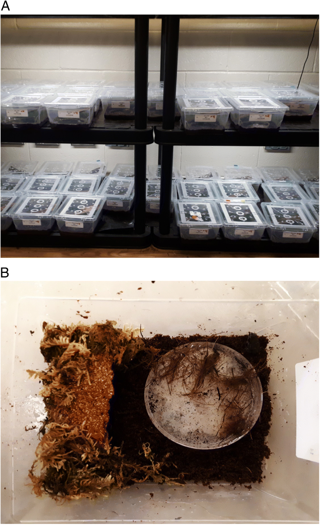
For each clutch, water was maintained at 21 °C and a pH of 7.5. All water conditions were established in accordance to the parameters recorded in the wild across three seasons of fieldwork (Bienentreu 2019). The photoperiod was set to 14:10 h light–dark hours using full spectrum 4000 k LED light bars (1 per 10 experimental units). A 75% water change with dechlorinated aged water (21 °C) was conducted on a 6-d basis, and two air diffusers were added to each tub for oxygenation. Tadpoles were fed standard dried tadpole micropellets (ZooMed, Zoo Med Laboratories Inc., San Luis Obispo, California, USA) on a 2-d basis: 30 mg/tadpole for week 1, 60 mg/tadpole for week 2, and 120 mg/tadpole for week 3 and until metamorphosis. This amount of food corresponds to an intermediate level of resources, appropriate for not overfeeding the larvae (Murray 1990). Loose coconut fibres were provided as environmental enrichment. Dead tadpoles were immediately removed and examined for typical signs of ranaviral disease (e.g., edema of ventral body and extremities; dermal ulcerations and hemorrhages; see Miller et al. 2015). Upon tadpoles reaching Gosner stage 32 (Gosner 1960), 10 tadpoles per clutch were haphazardly chosen and euthanized via immersion in 6% MS222 solution. We pooled tissue samples per clutch and screened for ranavirus using quantitative polymerase chain reaction (qPCR). All samples tested negative. Subsequently, 30 tadpoles from each of the four clutches were separated into 120 individual plastic tubs (2.1 L food grade, Fig. 1A), filled with 1.6 L dechlorinated aged water, and monitored daily. We randomly redistributed the plastic tubs on our shelving units every 3 d to eliminate any effects due to specific placement in the setup (e.g., light intensity and microtemperature differences). After reaching Gosner stage 40, each tadpole was provided with a floating cork platform to allow for resting and to prevent drowning. The amount of water in each tub was gradually decreased to 0.2 L as each animal progressed to Gosner stage 45. A perforated lid was added to each tub to prevent the animals from escaping. To prevent fouling of the water, we stopped feeding during metamorphic climax (Gosner stages 42–45) as animals temporarily stop eating during these stages. All 120 tadpoles completed metamorphosis within the first week of July 2018 (mean time hatch to metamorphosis: 49 ± 3 d). All water was then removed from the enclosures, and coconut fibre substrate (Eco Earth, Zoo Med Laboratories Inc., San Luis Obispo, California, USA), a small amount of forest moss (Exo Terra, Rolf C. Hagen Inc., Montreal, Quebec, Canada) to maintain moisture, and a 100-mm petri dish filled with dechlorinated water were added to each tub. Natural cork sheets were used to create a shelter but arranged such that animals could not hide out of sight (Fig. 1B). Frogs were held at 21 °C day and 18 °C night temperature, and the photoperiod was set to 14:10 h light–dark cycles. Approximately 15 flightless fruit flies (Drosophila melanogaster) were provided every 2 d for the first 14 d. We then introduced bigger fruit flies (D. hydei), in variation with house crickets (Acheta domesticus) and banded crickets (Gryllus sigillatus), as well as mealworms (Tenebrio molitor) as food sources. Individuals were either fed 20 fruit flies, two small crickets, or two small mealworms on a 2-d basis. We used Exo Terra Calcium and Multi Vitamin Powder in a 1:1 ratio to dust insects and provide additional nutrient and mineral supply. We used Repashy Superfly Fruit Fly Culture Medium (Repashy Ventures Inc., Oceanside, California, USA) as well as Fluker’s Orange Cube Complete Cricket Diet (Fluker’s Cricket Farm Inc., Port Allen, Louisiana, USA) to maximize the nutritional value of the insects.
Fig. 1.
Fig. 1. Experimental setup. (A) Shelving system containing experimental units equipped with full spectrum 4000 k LED light bars (1 per 10 experimental units). (B) Experimental unit close-up (coconut fibre substrate, forest moss to maintain moisture, 100-mm petri dish filled with dechlorinated aged water, natural cork sheet as shelter).

Experimental exposures

The exposure trials were conducted with wood frogs that were 45–50 d postmetamorphosis (Gosner stage 46: Gosner 1960). A total of 100 frogs (25 from each of the four clutches) were randomly selected and equally assigned to the five treatments (20 frogs per treatment). Within each treatment, frogs had a snout-vent length (SVL) of 22.5 ± 2.5 mm and a weight of 1.5 ± 0.5 g. To simulate a natural route of infection, we conducted a 2 h individual water bath exposure (Gray et al. 2009a; Bayley et al. 2013) in 200 mL glass jars, containing 15 mL water with either 102.97 PFU/mL (low titre) or 103.97 PFU/mL (high titre) of the appropriate virus isolate suspended in RPMI medium. Individuals in the negative control treatment were challenged with RPMI medium only, following the same procedure. The media volume used was sufficient to guarantee full coverage of the ventral and dorsal surfaces of the frogs without the possibility of drowning. All individuals were monitored twice daily for morbidity and gross signs of ranavirosis (e.g., Gray et al. 2009a; Miller et al. 2015). Activity level and feeding behaviours were also recorded immediately each time on a 3-level scale (activity: 0 = inactive, 1 = passive–sheltered, 2 = active–outside; feeding: 0 = no reaction to food, 1 = interest–approach, 2 = feeding). All frogs were measured and weighed every 3 d, and thoroughly examined for external pathology consistent with ranavirosis (e.g., edema of ventral body and extremities, dermal ulcerations and hemorrhages; see Miller et al. 2015) to avoid inadvertently missing transient indications of infection. To minimize stress, the handling time for each animal was reduced to a routine of approximately 20 s. This routine was: (i) place container (with frog) on a scale, (ii) transfer frog into new Ziploc bag, (iii) determine SVL and examine for external signs of infection, (iv) tare scale, (v) place frog back into container, and (vi) determine weight. All dead frogs were removed from their tanks and necropsies were immediately performed: frogs were thoroughly examined for external and internal signs of ranavirosis (internal signs: hemorrhages and necrosis, in particular in liver and spleen; see Miller et al. 2015). Liver sections were removed and stored individually in 1.5-mL microcentrifuge tubes (Eppendorf) filled with 95% ethanol and frozen at −70 °C until processed for qPCR. The remainder of each carcass was placed in either 10% buffered formalin for histology or RNAlater for further analysis. The experiment was terminated 40 d postexposure, a sufficient duration for morbidity and mortality to occur (Cullen et al. 1995; Cunningham et al. 2007a). All remaining frogs in the virus treatments and the control group were individually euthanized by immersion in 6% MS222 solution and subjected to postmortem procedures as described above. The experiment was conducted under Laurentian University Animal Care Protocol #6013781.

qPCR-based ranavirus quantification

Tissue samples were assessed for ranavirus following the protocol for quantitative PCR (qPCR) described by Leung et al. (2017), and viral loads were quantified as described by Hoverman et al. (2010). DNA extraction from liver sections was performed using Qiagen DNEasy Blood and Tissue kits according to manufacturer specifications (QIAGEN Inc., Valencia, California, USA). For quantification of genomic DNA, a Synergy H1 Hybrid Multi-Mode Reader (BioTek, Winooski, Vermont, USA) and a Quant-iT dsDNA BR Assay Kit (Invitrogen Corp., Carlsbad, California, USA) were used. For qPCR we used a Mx3005P qPCR System (Agilent Technologies, Santa Clara, California, USA). The qPCR mixture contained: 250 ng of template DNA, 10 μL TaqMan Universal PCR Master Mix 2X (Thermofisher Scientific), 1 μL forward primer MCPRV_F–5GTCCTTTAACACGGCATACCT3 (10 μM), 1 μL reverse primer MCPRV_R–5ATCGCTGGTGTTGCCTATC3 (10 μM), and 0.05 μL TaqMan probe MCP_NFQ–5TTATAGTAGCCTRTGCGCTTGGCC3 (100 μM), as well as PCR-grade water in the appropriate amount to reach the final reaction volume of 20 μL. Samples were run in duplicate at 50 °C for 2 min, 95 °C for 10 min, and 50 cycles of 95 °C for 15 s and 60 °C for 30 s. Individuals were considered positive if both duplicates showed a clear amplification (e.g., surpassing the respective cycle threshold). If only one of the two runs showed amplification, a third run was conducted, to either confirm or dismiss previous results. Each 96-well plate included a no-template control (DNA grade water) and a serial dilution of a known quantity of cultured FV3 (106–101 PFU/mL) to create standard curves with precise fit (R2 = >0.95). The primers used target a consensus sequence within major capsid protein that is shared among the majority of amphibian associated Ranavirus isolates (FV3, GenBank AY548484, Tan et al. 2004; tiger frog virus, GenBank AF389451, He et al. 2002; Common midwife toad virus, GenBank JQ231222, Jancovich et al. 2010; Epizootic haematopoietic necrosis virus, GenBank FJ433873, Mavian et al. 2012), and allows for a high analytical sensitivity when used in combination with a TaqMan probe (Leung et al. 2017). Following Yuan et al. (2006), the standard curves were used to calculate viral load in copies/250 ng gDNA, as recommended by Gray and Chinchar (2015). Due to large standard deviations, viral loads are reported as log10 (copies/250 ng gDNA).

Histopathology

At the end of the experiment (40-d postexposure) two individuals per treatment were assessed for lesions in liver tissue using histopathology. Both chosen individuals exhibited signs during the experiment, with one clearing the signs and the other exhibiting signs, until the end of the experiment. All frogs that died during the experiment, as well as two frogs of the control were also examined histologically. Tissues were routinely processed and embedded in paraffin, sectioned at 5 μm using a rotary microtome, and stained with hematoxylin and eosin. Sections were examined using a Leica D500 light microscope, with images captured using a Leica ICC50W camera and LAS software (V 4.5, Leica, Wetzlar, Hesse, Germany).

Statistical analyses

Statistical analyses were conducted using R v3.3.3 (R Core Team 2013) in R Studio V3.4.1 (RStudio Team 2016). We used χ2 tests to compare survival rates, infection rates, and presentation of gross pathology associated with ranavirus infections. Survival rates were compared across all treatments. whereas comparisons of infection and pathology rates did not include the control group. We conducted mixed-effect ANOVAs with clutch as a random effect to test for differences among virus treatments with respect to percentage growth (SVL and weight), activity level, mean viral loads, and feeding behaviour. We also conducted one-way ANOVAs to test for differences in the aforementioned response variables among the different clutches within and across treatments. Since the control group had no infected individuals, resulting in a mean and SD = 0, it was removed from the ANOVAs for viral loads. We used Tukey’s Honest Significant Difference test to detect differences among means for ANOVAs where the overall null hypothesis was rejected. We conducted a sensitivity analysis of the body condition data for each treatment to detect potentially influential outliers (R packages “broom”: Robinson et al. 2019, and “dplyr”: Wickham et al. 2019).

Results

Infection and mortality rates

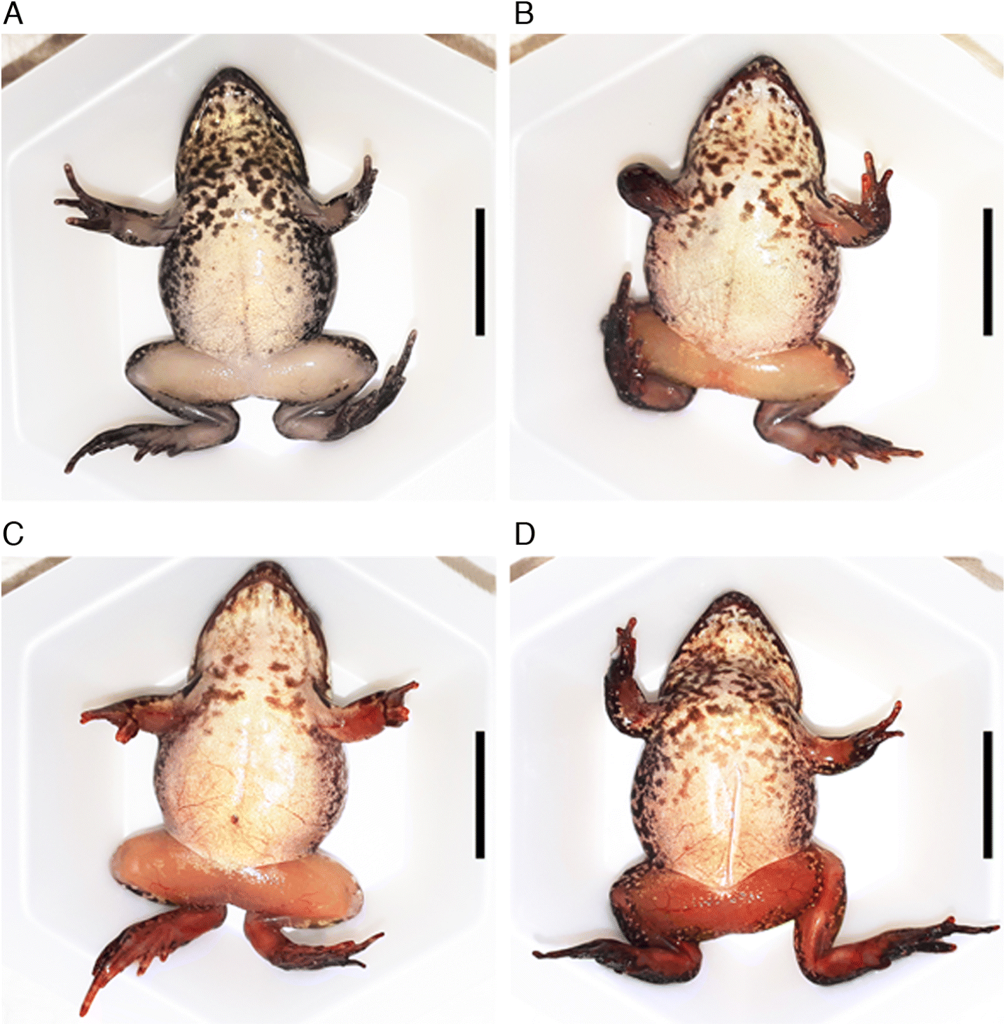
No frogs from the control treatment died, tested positive for FV3 via qPCR, or exhibited any gross pathology associated with ranavirosis (Table 1). Overall, we observed a 42% infection rate and a 5% ranavirus-related mortality in postmetamorphic wood frogs exposed in the different virus treatments (Table 1). Chi-squared tests did not show any significant differences in survival rates among all treatment groups. Infection rates, mortality due to ranavirosis, or gross signs of infection did not differ significantly among the virus treatments. Infection rates followed a dose-dependent pattern in both the WT and KO FV3 challenges although the trends were not statistically significant (Table 1). Two individuals in the high-dose WT FV3 group died between 8 and 10 d postexposure, and one individual died in each KO FV3 treatment, 8 and 11 d postexposure. We observed severe external haemorrhaging in these animals 1–2 d before death, consistent with gross pathology associated with ranavirosis in other studies (e.g., Gray et al. 2009a; Miller et al. 2015) and internal haemorrhages, particularly in liver and kidney tissue. Four other individuals died within the last 10 d of the experiment, one frog in each virus treatment. All four of these frogs tested negative for ranavirus via qPCR, but two of the four individuals exhibited signs of infection (e.g., haemorrhages, erythema) 2–3 d prior to death (WT FV3 low and KO FV3 high treatment). While performing postmortem examinations, we did not observe any gross external or internal signs of infection in these individuals. Starting at approximately day 7 postexposure, individuals in the virus treatments started to show gross signs of ranavirosis, such as diffusely congested blood vessels and erythema, primarily on the extremities, toes and fingers, as well as on the anterior ventral body surface (Figs. 2A2D). Such signs were present in 79% and 94% of the individuals exposed to the WT FV3 treatments (low and high, respectively) as well as 72% and 83% in the KO FV3 treatments (low and high, respectively). At the end of the study, 87% and 81% of individuals previously exhibiting lesions in the low and high WT FV3 infection groups, respectively, and 85% and 73% of the low and high KO FV3 groups, respectively, no longer had any observable external signs of disease (Table 1). Mean viral loads at the end of the experiment differed in a dose-dependent pattern, although the trend was not statistically significant (Table 2).
Table 1.
Table 1. Survival and proportion of qPCR positive individuals (alive and dead) in postmetamorphic wood frogs 40 d postexposure to WT FV3 and KO FV3 in water bath, as well as overall frequency of individuals (alive and dead) exhibiting heamorrhages during the experiment and number of individuals that cleared heamorrhages prior to death/euthanasia.
 WT lowWT highKO lowKO highControldfχ2p
Survival19/20 (95%)17/20 (85%)18/20 (90%)18/20 (90%)20/20 (100%)40.110.991
Alive PCR(+)7/19 (37%)9/17 (53%)4/18 (22%)10/18 (56%)0/20 (0%)32.800.424
Dead PCR(+)0/1 (0%)2/3 (67%)1/2 (50%)1/2 (50%)N/A32.010.572
Hem alive15/19 (79%)16/17 (94%)13/18 (72%)15/18 (83%)0/20 (0%)30.320.956
Hem cleared alive13/15 (87%)13/16 (81%)11/13 (85%)11/15 (73%)N/A30.330.954
Hem dead1/1 (100%)2/3 (67%)1/2 (50%)2/2 (100%)N/A30.670.881
Hem cleared dead1/1 (100%)0/2 (0%)0/1 (0%)1/2 (50%)N/A3N/AN/A

Note: Respective χ2 and p values included. Viral titres: 102.97 PFU/mL (low) or 103.97 PFU/mL (high). qPCR, quantitative polymerase chain reaction; WT, wild-type; FV3, Frog virus 3; KO, knock-out; df, degrees of freedom; hem, hemorrhages; PFU, plaque forming units; N/A, Not applicable.

Table 2.
Table 2. Relative growth (length and weight gain; mean (± SD) viral load, activity level and feeding behaviour for postmetamorphic wood frogs (Rana sylvatica) after bath exposure to WT FV3 and KO FV3, 40 d postexposure.
 WT lowWT highKO lowKO highControldfFp
Length gain %19.11 ± 6.1622.47 ± 5.9314.5 ± 6.112.83 ± 5.9518.75 ± 5.054, 877.73<0.001
Weight gain %57.16 ± 27.0165.65 ± 29.6452.22 ± 22.7343.78 ± 20.9559.1 ± 24.624, 871.870.123
AVL all ind0.86 ± 1.181.3 ± 1.290.5 ± 0.971.54 ± 1.52N/A3, 681.080.364
AVL pos ind2.34 ± 0.322.45 ± 0.352.26 ± 0.092.78 ± 0.75N/A3, 260.750.534
AVL dead indN/A8.37 ± 0.475.72 ± 08.84 ± 0N/A2, 11.520.498
Activity level1.24 ± 0.131.41 ± 0.141.23 ± 0.091.3 ± 0.161.19 ± 0.154, 877.32<0.001
Feeding behaviour1.38 ± 0.201.42 ± 0.241.37 ± 0.181.44 ± 0.171.33 ± 0.224, 870.830.512

Note: Mean viral loads are shown as log (copies/250 ng gDNA). For each response variable, respective df, F and p are shown. Viral titres: 102.97 PFU/mL (low) or 103.97 PFU/mL (high). WT, wild-type; FV3, Frog virus 3; KO, knock-out; df, degrees of freedom; AVL, average viral load; ind, individuals; pos, positive; N/A, not applicable.

Fig. 2.
Fig. 2. Gross lesions of varying intensity observed in postmetamorphic wood frogs (Rana sylvatica) experimentally infected with ranavirus, 40 d postexposure: (A) no obvious signs (wild-type Frog virus 3 (WT FV3) low); (B) slight haemorrhages in fingers and minor erythema on the anterior ventral body surface (WT FV3 high); (C) moderate erythema in extremities as well as haemorrhages in fingers and toes (knock-out (KO) FV3 high); and (D) severe erythema in extremities and anterior ventral body surface, diffusely congested blood vessels, as well as haemorrhages in fingers and toes (WT FV3 high). Scale bar = 10 mm.

Growth, activity, and behaviour

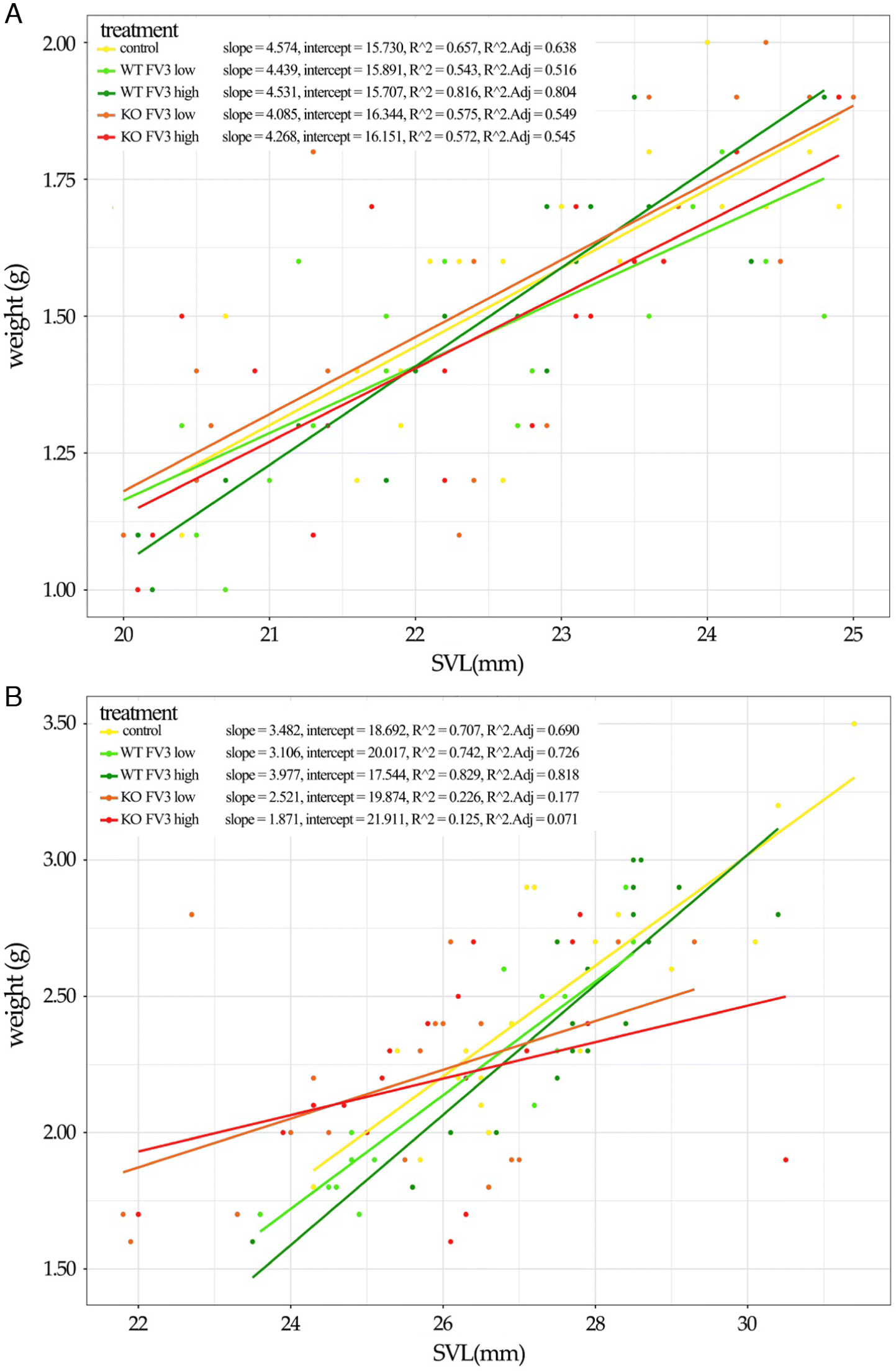
There were no significant differences among treatments with respect to feeding behaviour or relative weight gain among the treatments. Similarly, there were no statistical differences among egg clutches for any response variable (Supplementary Material S1-I). We observed significant variation in relative length gain among treatments (F4,87 = 7.73, p < 0.001; Table 2). Post hoc tests revealed that the length gain in the KO FV3 high treatment was significantly lower than the WT FV3 and the control treatments, with the largest difference relative to the high-dose WT FV3 group (p < 0.001). Overall, the two KO FV3 infection groups exhibited a 11% lower relative length increase compared with the control group (Table 2; Fig. 3), whereas the WT FV3 treatments showed a 2% higher length gain than the control animals. Activity level of individuals also showed significant variation among treatments (F4,87 = 7.32, p < 0.001; Table 2). Post hoc tests indicated that individuals in the WT FV3 high-dose treatment were significantly more active than those in the WT FV3 low treatment (p < 0.01) or the control group (p < 0.001). The KO FV3 high treatment showed significantly lower activity levels relative to all other treatment groups (p < 0.05), whereas the KO FV3 low-dose treatment showed significant differences to the WT FV3 high-dose group only (p < 0.01). A sensitivity analysis of the body condition data for each treatment did not show any significant outliers (Supplementary Material S1-II).
Fig. 3.
Fig. 3. Comparison of body condition (regression of length~weight) of postmetamorphic wood frogs (Rana sylvatica) water-bath exposed to ecologically relevant doses of (wild-type Frog virus 3 (WT FV3) and knock-out (KO FV3), at the start (A) and end (B) of the experiment (including respective slope, intercept, R2, and adjusted R2). SVL, snout-vent length.

Histopathology

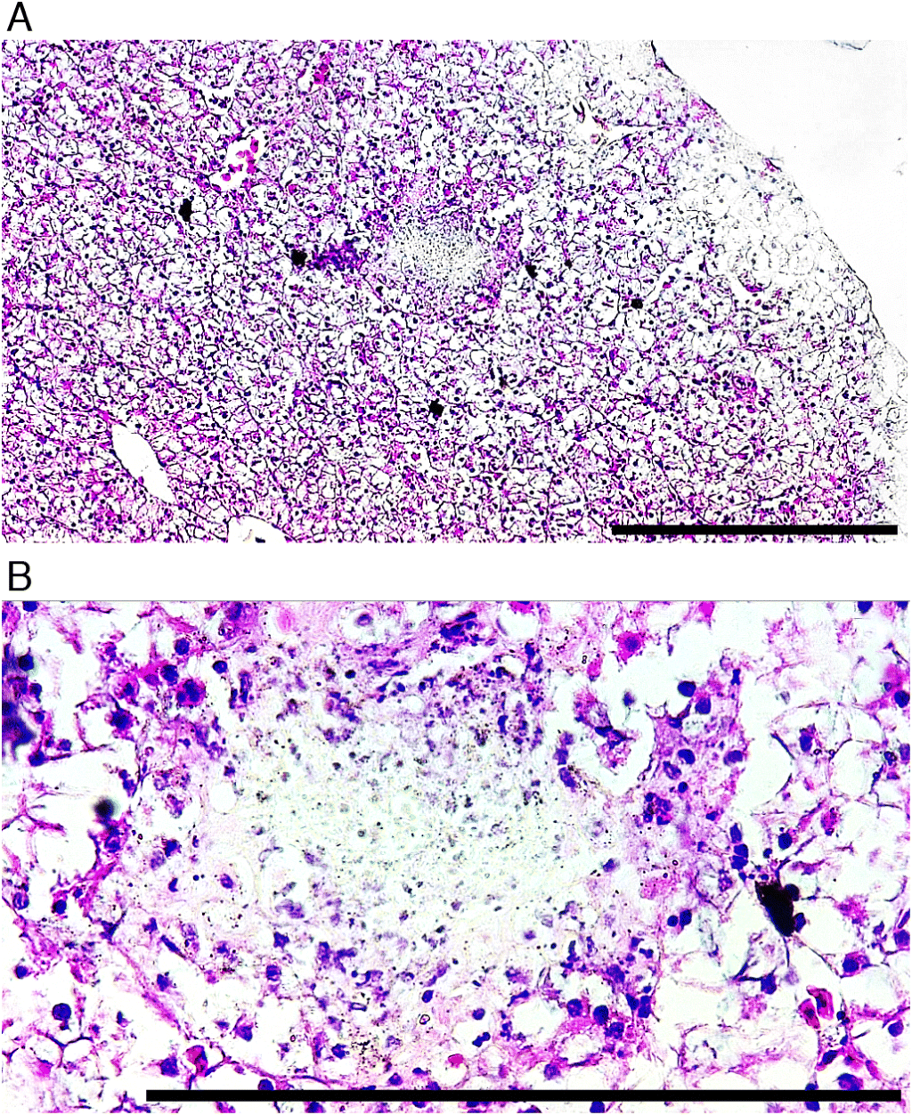
All four asymptomatic individuals showed no lesions, whereas the four symptomatic individuals across the treatments exhibited hepatocellular necrosis with the loss of the normal trabecular pattern of liver tissue (Figs. 4A and 4B). All four symptomatic individuals who died within the first 11 d of the experiment also exhibited aforementioned pathology. No histopathologic signs indicative for ranavirus infection were observed in liver tissue of the individuals who died in the last 10 d of the experiment or in the control group.
Fig. 4.
Fig. 4. Hematoxylin/eosin stained liver sections of postmetamorphic wood frogs (Rana sylvatica) experimentally infected with ranavirus, 40 d postexposure. Multifocal hepatocellular necrosis, (wild-type Frog virus 3 (WT FV3) high (A); foci in detail (B), showing severe hepatocellular necrosis, associated with the abundance of cellular debris, WT FV3 high. Bars = 1000 μm.

Discussion

Ranavirus induced mortality was very low (5%) in postmetamorphic wood frogs when exposed to environmentally relevant concentrations of ranavirus via water bath, and only 42% of the frogs tested positive for ranavirus across the virus treatments at the end of the 40-d experiment. Infection rates and viral loads followed a dose-dependent pattern, but we did not observe any significant differences in mortality between the virus treatments. The observed times to death (8–11 d) were consistent with other studies on postmetamorphic individuals (e.g., Bayley et al. 2013; Forzán et al. 2015), and although there was abundant gross pathological evidence of infection within the first 2 weeks postexposure, most individuals showed no or minor signs of infection at the end of the trial.
The overall low infection and mortality rates may be a consequence of selection towards more resilient genetic lineages within the source population, induced by repeated exposure to the pathogen (Pearman and Garner 2005). The genetic composition of distinct populations may have significant impact on the outcomes of a ranavirus emergence (Echaubard et al. 2014), and a co-evolutionary history of the host immune system with the pathogen may have led to decreased susceptibility to re-introduced pathogens or strains thereof (Pearman and Garner 2005). All wood frogs used in our study were raised from four egg clutches collected from a natural population within Wood Buffalo National Park that showed low ranavirus prevalence and low viral loads in 2017 (Bienentreu 2019). However, ranavirus has been frequently detected in amphibians in the area (Bienentreu 2019), with two confirmed die-off events in 2017 at wetlands located 7 and 29 km away from the sampled population (Forzán et al. 2019) and mortality occurring among wood frog tadpoles within 3.5 km in 2009 (D.M. Schock, unpublished work). Additionally, viral isolate sequencing led to the identification of at least two different FV3-like viruses prevailing in this area (Grant et al. 2019; Vilaça et al. 2019). These observations, in combination with our experimental results, could indicate that the source population evolved partial immunity against FV3-like ranaviruses due to repeated exposures to the pathogen.
An alternative explanation for the observations made in our experiment may be linked to the immune status of the postmetamorphic individuals. Experimental studies conducted with different life stages of the model species Xenopus laevis identified significant differences in the antiviral responses mounted by tadpoles and adult frogs to FV3, presumably reflecting the morphological and immunological differences between pre- and postmetamorphic anurans (Grayfer et al. 2015b; Wendel et al. 2017). Gene expression studies showed that tadpoles exhibit considerably less robust and delayed anti-FV3 inflammatory gene responses relative to adults (Andino et al. 2012), although they have rather timely antiviral (type III IFN) responses to this pathogen (Grayfer et al. 2015a; Wendel et al. 2017). Further, postmetamorphic X. laevis are capable of clearing FV3 infection within one month after exposure (Gantress et al. 2003). Therefore, our results may be an underestimate of the actual infection rates and viral loads that occurred over the duration of the entire experiment. Frogs that persistently exhibited gross signs of infection until the end of the study had low viral loads but showed severe hepatocellular necrosis. Perhaps a considerably greater proportion of the frogs exposed to virus in our study became infected with FV3, but subsequently reduced FV3 levels in their liver tissues below the detection capabilities of the methods employed here. This possibility is consistent with our observations that 81% of the frogs that exhibited gross signs of infection in the first two weeks postexposure were apparently healthy by the end of the experiment, and only 42% tested positive for the virus. We chose to focus on liver tissues because the gastrointestinal system and, in particular, the liver are commonly affected in ranavirus infections (Forzán et al. 2015; Miller et al. 2015). However, histological evidence of FV3 infections in adult wood frogs indicates that the virus can also persist in bone marrow and organs of the lymphatic and urinary system (Forzán et al. 2015). All four animals (one per virus treatment) that died late in the study tested negative for FV3 in their livers via qPCR, but two of the four exhibited signs of infection in the first two weeks of the experiment. Some or all of these frogs may sustained tissue damage in other organs, ultimately resulting in their death, as found by Grayfer et al. (2014) using FV3 in X. laevis. We did not detect any histological lesions in liver tissues of these four animals, and unfortunately, we were unable to examine other tissues for lesions. Thus, it remains unknown whether FV3 was in other tissues of these four frogs.
FV3 infects a wide variety of cold-blooded vertebrates which possess a myriad of immune systems and broad differences in their respective antiviral immune responses. We know relatively little about the evolutionary origins of this pathogen and which of its aquatic vertebrate host immune system(s) this virus has coevolved with. Thus, it stands to reason that the FV3 immune evasion mechanisms are best suited for interacting with immune proteins of those species in the context of which the virus has evolved. For example, the FV3 vIF-2α is a better decoy substrate for the RNA-induced protein kinase R (PKR) of some species over others (Andino et al. 2015; Grayfer et al. 2015a). While the WT and KO FV3 clearly possess broad immune evasion differences in other amphibian species (Andino et al. 2015; Grayfer et al. 2015a), the fact that we did not see marked differences between WT and vIF-2α KO FV3 infections of wood frogs may indicate that the FV3 vIF-2α is less effective or compatible with the wood frog immune system in general and with these frogs PKR in particular. In fact, R. sylvatica is considerably resistant to this FV3 possibly reflecting that FV3 did not coevolve with this species or related ones and is thus less effective at infecting it. In general, the magnitudes of FV3 loads do not as clearly correlate with infection outcomes, as is commonly seen with other viral infections (Grayfer et al. 2014). For example, infected adult X. laevis have been shown to hold considerably higher viral loads than tadpoles (Grayfer et al. 2015a; Wendel et al. 2017), even though tadpoles typically succumb to ranavirosis (Landsberg et al. 2013; Reeve et al. 2013; Grayfer et al. 2014). Since the wood frogs used in our experiment experienced low mortality and signs of ranavirosis are often exhibited even at low titers (Grayfer et al. 2014), the differences in the viral loads would not necessarily be reflected in animal survival. Furthermore, viral loads were solely examined from liver tissue and no other organs were screened in this study. It has been shown that kidney and liver FV3 loads can differ in X. laevis tadpoles and that immune modulation has different effects on the FV3 loads in different tissues of these animals (Grayfer et al. 2015a). Thus, it is possible that the replication of vIF-2α KO FV3 may be more immunologically restricted in distinct tissue(s) other than the liver. If this is the case in wood frogs, FV3 loads of WT and KO infected animals could have differed in organs other than the liver.
Notably, wood frogs exposed to the KO FV3 showed lower activity and decreased growth, as well as slightly higher mean viral loads in the high-dose treatment, compared with wood frogs exposed to the WT FV3. These effects are potentially due to energetically costly specific immune responses. Vertebrate antiviral defenses are highly dependent on IFN cytokine-mediated immunity. Recent in vitro studies suggest that the FV3 vIF-2α gene product is crucial to counteracting the host IFN-induced antiviral states (Grayfer et al. 2015a). Andino et al. (2015) showed that the KO FV3 exhibited reduced replication in vitro in the immuno-competent X. laevis A6 kidney epithelial cell line and elicited less pronounced type I and III IFN responses in A6 cultures, compared with the wild type FV3. Interestingly, when the A6 cell IFN responses elicited by the KO FV3 were reassessed as a function of the viral loads in those cultures, the KO FV3 elicited proportionately greater IFN responses than the WT FV3 (L. Grayfer, unpublished work). This, in turn, confirms that despite deletions in its N-terminal region, the FV3 vIF-2α gene product is crucial to dampening the frog host IFN responses (Grayfer et al. 2015a). While the functional significance of the truncation remains to be fully understood, our results indicate that this FV3 gene plays an important role in evading the host antiviral IFN response. We thus hypothesize that the lower activity and the decreased growth we observed in wood frogs infected with the vIF-2α KO FV3 reflect their more pronounced, and thus presumably more energetically costly, cytokine responses relative to animals infected with WT FV3. Conversely and as proposed above, the lack of significant viral load and mortality differences between WT- and vIF-2α KO FV3-infected animals could be attributed to less effective (albeit some) WT FV3 vIF-2α–host immune system (PKR) interactions. Alas, these hypotheses remain speculative in the absence of comprehensive immunological data.
We chose water-bath exposure, because short time exposures to water containing virus particles shed by other (possibly asymptomatic) infected individuals is the most likely route of infection, since postmetamorphic individuals only infrequently visit wetlands (Regosin et al. 2003). Direct transmission between individuals (as shown in Cullen et al. 1995; Picco et al. 2007) likely only occurs at higher rates during breeding aggregations in the spring. The long-term persistence of ranaviruses in host communities has been linked to competent amphibian reservoirs (Brunner et al. 2004). Field studies as well as experimental exposures to different ranavirus isolates have shown that individuals may sustain sublethal infections (Pearman et al. 2004; Gray et al. 2007; Greer et al. 2009; Miller et al. 2009; Hoverman et al. 2011) and can shed sufficient amounts of ranavirus virions to infect other individuals (103–104 PFU/mL: Brunner et al. 2004; Robert et al. 2005; Rojas et al. 2005). Furthermore, overwintering tadpoles and paedomorphic salamanders can act as reservoir in aquatic habitats (Brunner et al. 2004; Gray et al. 2009a), and several field studies have identified sublethal infected postmetamorphic amphibians, with only a minority exhibiting pathological signs (Australia and Europe: Cullen et al. 1995; Cunningham et al. 2007b; Ariel et al. 2009b; North America: Gray et al. 2009b; Hoverman et al. 2011; Vilaça et al. 2019). Our findings, as well as reports of re-occurring mass mortality events in other amphibian populations inhabiting semipermanent and ephemeral wetlands (Green et al. 2002; Petranka et al. 2007; Todd-Thompson 2010), further support a specific life-history stage reservoir theory. In such environments, suitable amphibian life-history stages are the most likely reservoirs for ranavirus persistence because virions will not remain active and viable for extensive period of times outside their host without a moist or even aquatic environment (Brunner et al. 2007; Nazir et al. 2012). Therefore, both larval and postmetamorphic amphibians can contribute to the persistence of the pathogen in the environment (Duffus et al. 2008).
Sample sizes in our experiment were based on other studies determining ranavirus susceptibility on species level (e.g., Hoverman et al. 2010; Sutton et al. 2014; Forzán et al. 2015), that showed that sample sizes of 20 individuals per treatment are sufficient to observe differences among treatments. The concentrations used were based on environmentally relevant concentrations of ranavirus virions (103–104 PFU/mL; Rojas et al. 2005; Gray et al. 2009a). Other studies using FV3 or FV3-like viruses with similar concentrations and bath exposure observed high mortality (97% and 100% for R. temporaria and R. servosus, respectively; Bayley et al. 2013; Sutton et al. 2014) highlighting the high variance in species susceptibility (Hoverman et al. 2010). Postmetamorphic wood frogs orally inoculated with similar FV3 concentrations showed 80%–100% mortality, and the odds of dying increased 23-fold for every tenfold increase in concentration, with death occurring approximately 15% earlier (Forzán et al. 2015). This suggests that mortality rates are concentration and route-specific, even within the same host species.
Identifying reservoirs that allow the pathogen to persist in the environment and facilitate its spread in animal communities remains a challenge of modern epidemiology. Using wood frogs, the most widely distributed amphibian species in North America (Martof 1970), we document important in vivo effects of infection of WT FV3 and KO FV3 in postmetamorphic wood frogs, contributing to the overall knowledge on infection in adult anurans and highlighting the function of the vIF-2α gene in ranavirus pathogenesis. Moreover, low viral loads in infected individuals underline the importance of high-sensitivity pathogen screening (Greer and Collins 2007), and provide evidence that even short duration exposures to environmentally relevant concentrations of ranavirus may cause sublethal infections in postmetamorphic wood frogs, indicating the role of this stage as a plausible reservoir for FV3 and possibly for ranavirus in general.
Acknowledgements

We respectfully acknowledge that egg collection was conducted on Treaty 8 Territory. Considerable logistical support was provided by Parks Canada staff Sharon Irwin and Katie McNab, Fort Smith, Northwest Territories, Canada. We thank the Xenopus laevis Research Resource for Immunobiology Resource at the University of Rochester, Rochester, New York, USA.

References

Andino FDJ, Chen G, Li Z, Grayfer L, and Robert J. 2012. Susceptibility of Xenopus laevis tadpoles to infection by the ranavirus Frog-Virus 3 correlates with a reduced and delayed innate immune response in comparison with adult frogs. Virology, 432(2): 435–443.
Andino FDJ, Grayfer L, Chen G, Chinchar VG, Edholm ES, and Robert J. 2015. Characterization of Frog virus 3 knockout mutants lacking putative virulence genes. Virology, 485: 162–170.
Ariel E, Nicolajsen N, Christophersen MB, Holopainen R, Tapiovaara H, and Jensen BB. 2009a. Propagation and isolation of ranaviruses in cell culture. Aquaculture, 294(3–4): 159–164.
Ariel E, Kielgast J, Svart HE, Larsen K, Tapiovaara H, Jensen BB, et al. 2009b. Ranavirus in wild edible frogs Pelophylax kl. esculentus in Denmark. Diseases of Aquatic Organisms, 85(1): 7–14.
Bayley AE, Hill BJ, and Feist SW. 2013. Susceptibility of the European common frog Rana temporaria to a panel of ranavirus isolates from fish and amphibian hosts. Diseases of Aquatic Organisms, 103(3): 171–183.
Bienentreu J-F. 2019. Epidemiology of ranaviruses in amphibian populations in the boreal forest of northwestern Canada. Ph.D. dissertation, Laurentian University, Sudbury, Ontario. 215 p.
Brunner JL, Schock DM, Davidson EW, and Collins JP. 2004. Intraspecific reservoirs: complex life history and the persistence of a lethal ranavirus. Ecology, 85(2): 560–566.
Brunner JL, Schock DM, and Collins JP. 2007. Transmission dynamics of the amphibian ranavirus Ambystoma tigrinum virus. Diseases of Aquatic Organisms, 77(2): 87–95.
Brunner JL, Barnett KE, Gosier CJ, McNulty SA, Rubbo MJ, and Kolozsvary MB. 2011. Ranavirus infection in die-offs of vernal pool amphibians in New York, USA. Herpetological Review, 42(1): 76–79.
Chen G, Ward BM, Kwang HY, Chinchar VG, and Robert J. 2011. Improved knockout methodology reveals that frog virus 3 mutants lacking either the 18K immediate-early gene or the truncated vIF-2α gene are defective for replication and growth in vivo. Journal of Virology, 85(21): 11131–11138.
Chinchar VG, Hyatt A, Miyazaki T, and Williams T. 2009. Family Iridoviridae: poor viral relations no longer. In Lesser known large dsDNA viruses. Edited by JL Van Etten. Springer, Berlin, Heidelberg, Germany. pp. 123–170.
Chinchar VG, Hick P, Ince IA, Jancovich JK, Marschang R, Qin Q, et al. 2017. ICTV virus taxonomy profile: Iridoviridae. Journal of General Virology, 98(5): 890–891.
Converse KA, and Green DE. 2005. Diseases of tadpoles. In Wildlife diseases: landscape epidemiology, spatial distribution, and utilization of remote sensing technology. Edited by SK Majumdar. The Pennsylvania Academy of Science. pp. 72–88.
Cullen BR, Owens L, and Whittington RJ. 1995. Experimental infection of Australian anurans (Limnodynastes terraereginae and Litoria latopalmata) with Bohle iridovirus. Diseases of Aquatic Organisms, 23(2): 83–92.
Cunningham AA, Hyatt AD, Russell P, and Bennett PM. 2007a. Experimental transmission of a ranavirus disease of common toads (Bufo bufo) to common frogs (Rana temporaria). Epidemiology and Infection, 135(7): 1213–1216.
Cunningham AA, Hyatt AD, Russell P, and Bennett PM. 2007b. Emerging epidemic diseases of frogs in Britain are dependent on the source of ranavirus agent and the route of exposure. Epidemiology and Infection, 135(7): 1200–1212.
Duffus ALJ, Pauli BD, Wozney K, Brunetti CR, and Berrill M. 2008. Frog virus 3-like infections in aquatic amphibian communities. Journal of Wildlife Diseases, 44(1): 109–120.
Duffus ALJ, Waltzek TB, Stöhr AC, Allender MC, Gotesman M, Whittington RJ, et al. 2015. Distribution and host range of ranaviruses. In Ranaviruses. Edited by MJ Gray and VG Chinchar. Springer. pp. 9–57.
Echaubard P, Little K, Pauli B, and Lesbarrères D. 2010. Context-dependent effects of ranaviral infection on Northern leopard frog life history traits. PLoS ONE, 5(10): e13723.
Echaubard P, Leduc J, Pauli B, Chinchar VG, Robert J, and Lesbarreres D. 2014. Environmental dependency of amphibian–Ranavirus genotypic interactions: evolutionary perspectives on infectious diseases. Evolutionary Applications, 7(7): 723–733.
Forzán MJ, Jones KM, Vanderstichel RV, Wood J, Kibenge FS, Kuiken T, et al. 2015. Clinical signs, pathology and dose-dependent survival of adult wood frogs, Rana sylvatica, inoculated orally with frog virus 3 Ranavirus sp., Iridoviridae. Journal of General Virology, 96(5): 1138–1149.
Forzán MJ, Bienentreu J-F, Schock DM, and Lesbarrères D. 2019. Multi-tool diagnosis of an outbreak of ranavirosis in amphibian tadpoles in the Canadian boreal forest. Diseases of Aquatic Organisms, 135(1): 33–41.
Gantress J, Maniero GD, Cohen N, and Robert J. 2003. Development and characterization of a model system to study amphibian immune responses to iridoviruses. Virology, 311(2): 254–262.
Gosner KL. 1960. A simplified table for staging anuran embryos and larvae with notes on identification. Herpetologica, 16(3): 183–190.
Granoff A, Came PE, and Rafferty KA Jr. 1965. The isolation and properties of viruses from Rana pipiens: their possible relationship to the renal adenocarcinoma of the leopard frog. Annals of the New York Academy of Sciences, 126(1): 237–255.
Grant SA, Bienentreu J-F, Vilaça ST, Brunetti CR, Lesbarrères D, Murray DL, et al. 2019. Low intraspecific variation of Frog virus 3 with evidence for novel FV3-like isolates in central and northwestern Canada. Diseases of Aquatic Organisms, 134(1): 1–13.
Gray MJ, and Chinchar VG. 2015. Introduction: history and future of ranaviruses. In Ranaviruses. Edited by MJ Gray and VG Chinchar. Springer. pp. 1–7.
Gray MJ, Miller DL, Schmutzer AC, and Baldwin CA. 2007. Frog virus 3 prevalence in tadpole populations inhabiting cattle-access and non-access wetlands in Tennessee, USA. Diseases of Aquatic Organisms, 77(2): 97–103.
Gray MJ, Miller DL, and Hoverman JT. 2009a. Ecology and pathology of amphibian ranaviruses. Diseases of Aquatic Organisms, 87(3): 243–266.
Gray MJ, Miller DL, and Hoverman JT. 2009b. First report of Ranavirus infecting lungless salamanders. Herpetological Review, 40(3): 316–319.
Grayfer L, Andino FDJ, and Robert J. 2014. The amphibian (Xenopus laevis) type I interferon response to frog virus 3: new insight into ranavirus pathogenicity. Journal of Virology, 88(10): 5766–5777.
Grayfer L, Andino FDJ, and Robert J. 2015a. Prominent amphibian (Xenopus laevis) tadpole type III interferon response to the frog virus 3 ranavirus. Journal of Virology, 89(9): 5072–5082.
Grayfer L, Edholm ES, Andino FDJ, Chinchar VG, and Robert J. 2015b. Ranavirus host immunity and immune evasion. In Ranaviruses. Edited by MJ Gray and VG Chinchar. Springer. pp. 141–170.
Green DE, Converse KA, and Schrader AK. 2002. Epizootiology of sixty-four amphibian morbidity and mortality events in the USA, 1996–2001. Annals of the New York Academy of Sciences, 969(1): 323–339.
Greer AL, and Collins JP. 2007. Sensitivity of a diagnostic test for amphibian Ranavirus varies with sampling protocol. Journal of Wildlife Diseases, 43(3): 525–532.
Greer AL, Berrill M, and Wilson PJ. 2005. Five amphibian mortality events associated with ranavirus infection in south central Ontario, Canada. Diseases of Aquatic Organisms, 67(1–2): 9–14.
Greer AL, Brunner JL, and Collins JP. 2009. Spatial and temporal patterns of Ambystoma tigrinum virus (ATV) prevalence in tiger salamanders Ambystoma tigrinum nebulosum. Diseases of Aquatic Organisms, 85(1): 1–6.
He JG, Lü L, Deng M, He HH, Weng SP, Wang XH, et al. 2002. Sequence analysis of the complete genome of an iridovirus isolated from the tiger frog. Virology, 292(2): 185–197.
Hoverman JT, Gray MJ, and Miller DL. 2010. Anuran susceptibilities to ranaviruses: role of species identity, exposure route, and a novel virus isolate. Diseases of Aquatic Organisms, 89(2): 97–107.
Hoverman JT, Gray MJ, Haislip NA, and Miller DL. 2011. Phylogeny, life history, and ecology contribute to differences in amphibian susceptibility to ranaviruses. EcoHealth, 8(3): 301–319.
Jancovich JK, Bremont M, Touchman JW, and Jacobs BL. 2010. Evidence for multiple recent host species shifts among the ranaviruses (family Iridoviridae). Journal of Virology, 84(6): 2636–2647.
Jensen BB, Holopainen R, Tapiovaara H, and Ariel E. 2011. Susceptibility of pike-perch Sander lucioperca to a panel of ranavirus isolates. Aquaculture, 313(1): 24–30.
Landsberg JH, Kiryu Y, Tabuchi M, Waltzek TB, Enge KM, Reintjes-Tolen S, et al. 2013. Co-infection by alveolate parasites and Frog virus 3-like ranavirus during an amphibian larval mortality event in Florida, USA. Diseases of Aquatic Organisms, 105(2): 89–99.
Lesbarrères D, Balseiro A, Brunner J, Chinchar VG, Duffus A, Kerby J, et al. 2012. Ranavirus: past, present and future. Biology Letters, 8(4): 481–483.
Leung WT, Thomas-Walters L, Garner TW, Balloux F, Durrant C, and Price SJ. 2017. A quantitative-PCR based method to estimate ranavirus viral load following normalisation by reference to an ultraconserved vertebrate target. Journal of Virological Methods, 249: 147–155.
Martof BS. 1970. Rana sylvatica. Catalogue of American Amphibians and Reptiles. Society for the Study of Amphibians and Reptiles, St. Louis, Missouri. pp. 86.1–86.4.
Mavian C, López-Bueno A, Balseiro A, Casais R, Alcamí A, and Alejo A. 2012. The genome sequence of the emerging common midwife toad virus identifies an evolutionary intermediate within ranaviruses. Journal of Virology, 86(7): 3617–3625.
Miller DL, Gray MJ, Rajeev S, Schmutzer AC, Burton EC, Merrill A, et al. 2009. Pathologic findings in larval and juvenile anurans inhabiting farm ponds in Tennessee, USA. Journal of Wildlife Diseases, 45(2): 314–324.
Miller DL, Pessier AP, Hick P, and Whittington RJ. 2015. Comparative pathology of ranaviruses and diagnostic techniques. In Ranaviruses. Edited by MJ Gray and VG Chinchar. Springer. pp. 171–208.
Murray DL. 1990. The effects of food and density on growth and metamorphosis in larval wood frogs (Rana sylvatica) from central Labrador. Canadian Journal of Zoology, 68(6): 1221–1226.
Nazir J, Spengler M, and Marschang RE. 2012. Environmental persistence of amphibian and reptilian ranaviruses. Diseases of Aquatic Organisms, 98(3): 177–184.
Pearman PB, and Garner TW. 2005. Susceptibility of Italian agile frog populations to an emerging strain of Ranavirus parallels population genetic diversity. Ecology Letters, 8(4): 401–408.
Pearman PB, Garner TW, Straub M, and Greber UF. 2004. Response of the Italian agile frog (Rana latastei) to a Ranavirus, frog virus 3: a model for viral emergence in naive populations. Journal of Wildlife Diseases, 40(4): 660–669.
Petranka JW, Harp EM, Holbrook CT, and Hamel JA. 2007. Long-term persistence of amphibian populations in a restored wetland complex. Biological Conservation, 138(3–4): 371–380.
Picco AM, Brunner JL, and Collins JP. 2007. Susceptibility of the endangered California tiger salamander, Ambystoma californiense, to ranavirus infection. Journal of Wildlife Diseases, 43(2): 286–290.
R Core Team. 2013. R: a language and environment for statistical computing. R Foundation for Statistical Computing, Vienna, Austria [online]: Available from R-project.org/.
Reeve BC, Crespi EJ, Whipps CM, and Brunner JL. 2013. Natural stressors and ranavirus susceptibility in larval wood frogs (Rana sylvatica). EcoHealth, 10(2): 190–200.
Regosin JV, Windmiller BS, and Reed JM. 2003. Terrestrial habitat use and winter densities of the wood frog (Rana sylvatica). Journal of Herpetology, 37(2): 390–394.
Robert J, Morales H, Buck W, Cohen N, Marr S, and Gantress J. 2005. Adaptive immunity and histopathology in frog virus 3-infected Xenopus. Virology, 332(2): 667–675.
Robinson D, Hayes A, Gomez M, Demeshev B, Menne D, Nutter B, et al. 2019. broom: convert statistical analysis objects into tidy tibbles. R package version 0.5.2 [online]: Available from cran.r-project.org/web/packages/broom/.
Rojas S, Richards K, Jancovich JK, and Davidson EW. 2005. Influence of temperature on Ranavirus infection in larval salamanders Ambystoma tigrinum. Diseases of Aquatic Organisms, 63(2–3): 95–100.
RStudio Team. 2016. RStudio: integrated development for R. RStudio, Inc., Boston, Massachusetts [online]: Available from rstudio.com/.
Schock DM, Bollinger TK, Gregory Chinchar V, Jancovich JK, and Collins JP. 2008. Experimental evidence that amphibian ranaviruses are multi-host pathogens. Copeia, 2008(1): 133–143.
Schock DM, Bollinger TK, and Collins JP. 2009. Mortality rates differ among amphibian populations exposed to three strains of a lethal ranavirus. EcoHealth, 6: 438–448.
Sutton WB, Gray MJ, Hardman RH, Wilkes RP, Kouba AJ, and Miller DL. 2014. High susceptibility of the endangered dusky gopher frog to ranavirus. Diseases of Aquatic Organisms, 112(1): 9–16.
Tan WG, Barkman TJ, Chinchar VG, and Essani K. 2004. Comparative genomic analyses of Frog virus 3, type species of the genus Ranavirus (family Iridoviridae). Virology, 323(1): 70–84.
Todd-Thompson M. 2010. Seasonality, variation in species prevalence, and localized disease for ranavirus in Cades Cove (Great Smoky Mountains National Park) amphibians. M.Sc. thesis, University of Tennessee, Knoxville, Tennessee. 43 p.
Vilaça ST, Bienentreu J-F, Brunetti CR, Lesbarrères D, Murray DL, and Kyle CJ. 2019. Frog virus 3 genomes reveal prevalent recombination between Ranavirus lineages and their origins in Canada. Journal of Virology, 93(20): e00765-19.
Vo NT, Guerreiro M, Yaparla A, Grayfer L, and DeWitte-Orr SJ. 2019. Class A scavenger receptors are used by frog virus 3 during its cellular entry. Viruses, 11(2): 93.
Watkins TB, and Vraspir J. 2005. Both incubation temperature and posthatching temperature affect swimming performance and morphology of wood frog tadpoles (Rana sylvatica). Physiological and Biochemical Zoology, 79(1): 140–149.
Wendel ES, Yaparla A, Koubourli DV, and Grayfer L. 2017. Amphibian (Xenopus laevis) tadpoles and adult frogs mount distinct interferon responses to the Frog Virus 3 ranavirus. Virology, 503: 12–20.
Wickham H, François R, Henry L, and Müller K. 2019. dplyr: a grammar of data manipulation. R package version 0.8.3 [online]: Available from cran.r-project.org/web/packages/dplyr/.
Yuan JS, Reed A, Chen F, and Stewart CN Jr. 2006. Statistical analysis of real-time PCR data. BMC Bioinformatics, 7(1): 85.

Supplementary material

Supplementary Material 1 (DOCX / 60 KB)

Information & Authors

Information

Published In

cover image FACETS
FACETS
Volume 5Number 1January 2020
Pages: 738 - 757
Editor: Ellen Ariel

History

Received: 3 January 2020
Accepted: 11 July 2020
Version of record online: 17 September 2020

Notes

This paper is part of a Collection titled “Ranavirus research: 10 years of global collaboration”.

Data Availability Statement

All relevant data are within the paper and Supplementary Material.

Key Words

  1. amphibian
  2. experimental exposure
  3. postmetamorphic
  4. ranavirus
  5. sublethal infection

Sections

Subjects

Authors

Affiliations

Joe-Felix Bienentreu
Department of Biology, Laurentian University, Sudbury, ON P3E 2C6, Canada
Leon Grayfer
Department of Biological Sciences, George Washington University, Washington, DC 20052, USA
Danna M. Schock
University Studies and Environmental Technology, Keyano College, Fort McMurray, AB T9H 2H7, Canada
Current address: Palustris Environmental, Athabasca, AB T9S 1H8, Canada.
Matthew Guerreiro
Department of Health Sciences, Wilfrid Laurier University, Waterloo, ON N2L 3C5, Canada
Melanie Mehes-Smith
Department of Biology, Laurentian University, Sudbury, ON P3E 2C6, Canada
Stephanie J. DeWitte-Orr
Department of Health Sciences, Wilfrid Laurier University, Waterloo, ON N2L 3C5, Canada
Jacques Robert
Department of Microbiology and Immunology, University of Rochester, Rochester, NY 14642, USA
Craig R. Brunetti
Department of Biology, Trent University, Peterborough, ON K9J 7B8, Canada
David Lesbarrères [email protected]
Department of Biology, Laurentian University, Sudbury, ON P3E 2C6, Canada

Author Contributions

J-FB, DMS, and DL conceived and designed the study.
J-FB, MG, and MM-S performed the experiments/collected the data.
All analyzed and interpreted the data.
SJD-O, JR, CRB, and DL contributed resources.
J-FB, LG, DMS, MG, SJD-O, JR, CRB, and DL drafted or revised the manuscript.

Competing Interests

The authors declare no conflict of interest. The funders had no role in the design of the study, in the collection, analyses, or interpretation of data, in the writing of the manuscript, or in the decision to publish the results.

Funding Information

This research was funded by NSERC to C.R.B. and D.L.; the Ontario Trillium Scholarship to J.F.B.; an NSF CAREER grant to L.G.; NIH and NSF to J.R.; NSERC Discovery grant and Ontario Ministry of Research, Innovation and Science (MRIS)’s Early Researcher Award (ERA) grant to S.D.-O.

Metrics & Citations

Metrics

Other Metrics

Citations

Cite As

Export Citations

If you have the appropriate software installed, you can download article citation data to the citation manager of your choice. Simply select your manager software from the list below and click Download.

Cited by

1. The wood frog (Rana sylvatica): An emerging comparative model for anuran immunity and host-ranavirus interactions
2. Prior induction of cellular antiviral pathways limits frog virus 3 replication in two permissive Xenopus laevis skin epithelial-like cell lines

View Options

View options

PDF

View PDF

Get Access

Media

Media

Other

Tables

Share Options

Share

Share the article link

Share on social media