Open access

The beneficial effect of natural antioxidants from olive oil with fig and date palm fruit extracts on biochemical and hematological parameters in rats treated with doxorubicin and γ-radiation

Publication: FACETS
9 July 2018

Abstract

The goal of this study was to determine the possible beneficial effect of olive oil (7 g/kg) with fig (1 g/kg) and date palm fruit (1 g/kg) extracts (OFD) on the toxicity hazards of doxorubicin (DOX) and (or) γ-radiation. The DOX-treated groups received doses of 2.5 mg/kg body weight via intravenous (IV) injection weekly for four consecutive weeks. Rats in the irradiated groups were exposed to whole-body γ-radiation with fractioned doses of 2 Gy weekly for four consecutive weeks. The OFD-treated groups received two weeks of pretreatment with OFD and daily supplementation via oral gavage during the experimental period. The DOX-treated and (or) irradiated groups showed decreases in the antioxidant parameters (reduced glutathione and nitric oxide) as well as increased lipid peroxidation products. In addition, we observed changes in the lipid profile parameters, lipid risk ratios, and hematological values (erythrocyte (RBC) count, hemoglobin (Hb) concentration, hematocrit (Hct) percentage, platelet count, and total and differential leukocyte (WBC) count) in these groups compared with the control rats. The administration of OFD to DOX-treated and (or) irradiated rats significantly ameliorated the oxidative stress markers, lipid profile, risk ratios, and hematological parameters. In conclusion, OFD could be used synergistically to decrease the negative side effects of chemotherapy and radiotherapy.

Introduction

Oxidative stress refers to the various harmful processes resulting from an imbalance caused by the excessive formation of reactive oxygen species (ROS) and the limited antioxidant defenses of cells (Turrens 2003). Doxorubicin (DOX) and (or) γ-radiation treatments are known to induce oxidative stress through the generation of ROS, which causes an imbalance in antioxidant activity and ultimately results in cell death (Elsadek et al. 2017; Fathy et al. 2017a). ROS can react with biological molecules and destroy the structure of cells (Baatout et al. 2004). They are often responsible for protein denaturation, lipid peroxidation, and impaired enzyme activity (Karbowink and Reiter 2000; Fathy et al. 2017a). DOX, also called adriamycin, is a potent antibiotic that is widely used for the treatment of different solid and hematopoietic tumors. However, in addition to its anti-tumoricidal activity, it has several well-known side effects that include chronic and irreversible toxicity (Asmis et al. 2005; Patil et al. 2008).
The chronic administration of plant extracts might augment the major cellular endogenous antioxidants, and is identified as a promising approach to combat oxidative stress (Bashandy et al. 2014; Fathy et al. 2017b). The major benefit of the Mediterranean diet is its high level of antioxidants derived from fruits and vegetables, including olive oil, figs, and date palm fruits, which contribute antioxidant vitamins, minerals, flavonoids, and polyphenol content (Solomon et al. 2006). In addition, mixed plant extracts showed a higher diversity of polyphenols resulting in greater stability and bioaccessibility of antioxidants compared with a single fruit extract (Bashandy et al. 2014; Kamiloglu et al. 2014; Fathy et al. 2018).
Our study aimed to investigate the protective synergistic effects of olive oil with fig and date palm fruit extracts on the toxicity hazards of DOX- and (or) γ-radiation-induced oxidative stress in Wistar albino rats.

Materials and methods

Ethics statement

All animals in our study were handled in accordance with the ethical guidelines for investigations using laboratory animals and complied with the guide for the care and use of laboratory animals (Institute of Laboratory Animal Resources 1996). The study was also approved by an independent ethics committee of the National Research Center, Egypt.

Experimental animals and study design

We used 120 male Wistar albino rats (150–170 g body mass). The rats were obtained from the Egyptian Holding Company for Biological Products and Vaccines (VACSERA, Giza, Egypt) and allowed to acclimatize in the experimental laboratory for two weeks. Rats were then divided into eight groups (n = 15 rats) according to the treatment and the requirements of the experiment. The rats were maintained under standard laboratory conditions at the animal center, Faculty of Pharmacy, Al-Azhar University, Cairo, Egypt. They were kept in a temperature-controlled environment (20–25 °C) and 50%–60% relative humidity with an alternating 12 h light:dark cycle. Five rats were placed into each cage and provided with standard diet pellets and drinking (tap) water ad libitum throughout the experimental period.
Group I (Control): Rats of this group were neither treated nor irradiated and were provided with standard diet pellets and drinking (tap) water ad libitum during the experiment (six weeks).
Group II (OFD): Rats of this group were administered extra virgin olive oil (7 g/kg) and freshly prepared fig (1 g/kg) and date palm fruit (1 g/kg) extracts daily via oral gavage for six weeks.
Group III (DOX): Rats of this group received DOX in doses of 2.5 mg/kg body weight via intravenous (IV) injection weekly for four consecutive weeks (cumulative doses of 10 mg/kg body weight).
Group IV (R): Rats of this group were exposed to whole-body γ-radiation with fractioned doses of 2 Gy every week for four consecutive weeks (up to 8 Gy total doses).
Group V (DOX-R): Rats of this group were irradiated following 20 h of DOX injection on the same schedule as Groups III and IV.
Group VI (OFD-DOX): Rats of this group were treated with OFD (for two weeks prior to DOX dosing and during the four weeks of treatment during the experiment) and injected with DOX on the same schedule as Group III.
Group VII (OFD-R): Rats of this group were treated with OFD (for two weeks prior to irradiation and during the four weeks of treatment during the experiment) and irradiated on the same schedule as Group IV.
Group VIII (OFD-DOX-R): Rats of this group were treated with OFD (for two weeks prior to DOX and R treatment and during the four weeks of treatment during the experiment) and irradiated following 20 h of DOX injection on the same schedule as Groups III and IV.

DOX

Adricin® (doxorubicin hydrochloride) vials were obtained from EIMC United Pharmaceuticals, Cairo, Egypt. Rats in our study were dosed with 2.5 mg/kg body weight of DOX via IV injection weekly for four consecutive weeks, for a cumulative dose of 10 mg/kg body weight.

Irradiation (R)

We used the Canadian Gamma cell-40 (137Cs) facility housed at the National Center for Radiation Research and Technology (NCRRT), Cairo, Egypt for irradiation treatment. Rats in the R-treated groups were exposed to whole-body γ-irradiation with fractioned doses (2 Gy every week for four weeks, up to 8 Gy cumulative doses). The dose rate at the time of the experiment was 0.45 Gy/min and the time of exposure was 4.44 min exactly.

Extra virgin olive oil (Olea europaea L., Family Oleaceae)

Monumental brand extra virgin olive oil was procured from the Grup Pons company (Lleida, Spain). The purchased extra virgin olive oil density was 920 g/L and the selected olive oil dose was 7.6 mL/kg (7 g/kg) body weight (Bashandy et al. 2014). The extra virgin olive oil was provided to the rats in the treatment group via oral gavage.

Fig fruit extract (Ficus carica L., Family Moraceae)

Dried ripe fig fruits were procured from Kafoods Ltd. (Istanbul, Turkey). The fig fruits were cut into small pieces, dried, and coarsely ground using an electrical device. The powdered material was soaked in five times its volume of 80% ethanol for 72 h with occasional shaking. The soaked material was filtered through fine filter paper, then subjected to evaporation under reduced pressure on a rotary evaporator until it dried (Gilani et al. 2008). Each rat in the treatment group received crude fig fruit extract at a concentration of 1 g/kg body weight (equivalent to about three figs) during the experimental period (six weeks) via oral gavage. The dose of the fig fruit extract was based on the recommended antioxidant dose of dry fig fruits for humans (Vinson et al. 2005) and converted to a suitable dose for albino rats (Reagan-Shaw et al. 2008).

Date palm fruit extract (Phoenix dactylifera L., Family Arecaceae)

The plant material was rendered free from soil and the date palm fruits were manually separated from the pits, and the flesh of the fruits was cut into small pieces, dried in an oven at 40 °C, and coarsely ground using an electrical device. The ground date palm fruits were added to ethanol (50%) (1:3 weight to volume) for 48 h in a refrigerator (4 °C) with continuous stirring (Al-Qarawi et al. 2005). The whole solution was ground, then centrifuged at 4 °C for 20 min at 1788g. The supernatant was collected and stored at −20 °C until used (Vayalil 2002). This suspension was given to rats via oral gavage, and each rat in the treatment group received crude date palm extract at a concentration of 1 g/kg body weight (equivalent to the flesh of seven dates) during the experimental period (six weeks). The dose of the crude date palm fruit extract was based on the recommended antioxidant dose of date palm fruits for humans (Vinson et al. 2005) and converted to a suitable dose for albino rats (Reagan-Shaw et al. 2008).

Biochemical study

Blood samples were collected at the end of the experiment from the retro-orbital venous plexus puncture of each animal (under anesthesia) using blood capillary tubes. One part (0.50 mL) of the blood sample was collected in ethylenediaminetetraacetic acid (EDTA) tubes for hematological study and the remaining sampled blood was left to clot at room temperature for 15 min. Sera were separated by centrifugation at 1006g at 20 °C for 15 min and the clear serum was extracted and kept frozen at −80 °C for use in the biochemical analyses. After blood sampling, the animals were sacrificed and the livers were isolated, quickly dissected out, and washed with isotonic ice-cold saline. A portion of each animal’s liver tissue was taken from all test groups. Each tissue sample was homogenized in ice-cold Tris–HCl lysis buffer (pH 7.4) containing 1% protease inhibitor cocktail (Cell Signaling Technology, Inc., Danvers, Massachusetts, USA) using Potter-Elvehjem rotor–stator homogenizer fitted with a Teflon pestle (Omni International, Kennesaw, Georgia, USA). The homogenates were centrifuged under cooling at 1006g for 20 min. All tissue samples were kept cold on a crushed ice at all times the preparation, and then supernatants were subsequently aliquot and stored at −80 °C until used for determination of hepatic thiobarbituric acid reactive substances (TBARS), reduced glutathione (GSH), and nitric oxide (NO) concentration.
TBARS were measured following the method of Yoshioka et al. (1979), NO was measured following the method of Montgomery and Dymock (1961), and GSH was measured following the method of Beutler et al. (1963). The serum lipid profile levels of triglycerides (TG), total cholesterol (TC), and high-density lipoprotein cholesterol (HDL-C) were estimated using kits from Elitech Diagnostic Co., France. The serum TG was determined according following the method described by Fossati and Prencipe (1982). The serum TC level was determined according to the method described by Allain et al. (1974). Serum HDL-cholesterol level was determined according to the method described by Burstein et al. (1970). Low-density lipoprotein cholesterol (LDL-C) was calculated according to the formula of Wieland and Seidel (1982).
The total number of erythrocytes (RBCs), total number of leukocytes (WBCs), differential leukocyte count, platelet count, hematocrit (Hct) percentage, and hemoglobin (Hb) concentration in the blood were estimated using a complete blood count (CBC) analyzer (Sinothinker sk9000, Shenzen, China).

Statistical analysis

Statistical analysis of the results was performed using statistical package for social sciences (SPSS) PC computer program (version 19, IBM Analytics, New York, New York, USA). All values were expressed as mean ± SE and the results were analyzed using one-way analysis of variance (ANOVA) test followed by least significant difference test (LSD) for multiple comparisons. Differences were considered statistically significant at p < 0.05.

Results

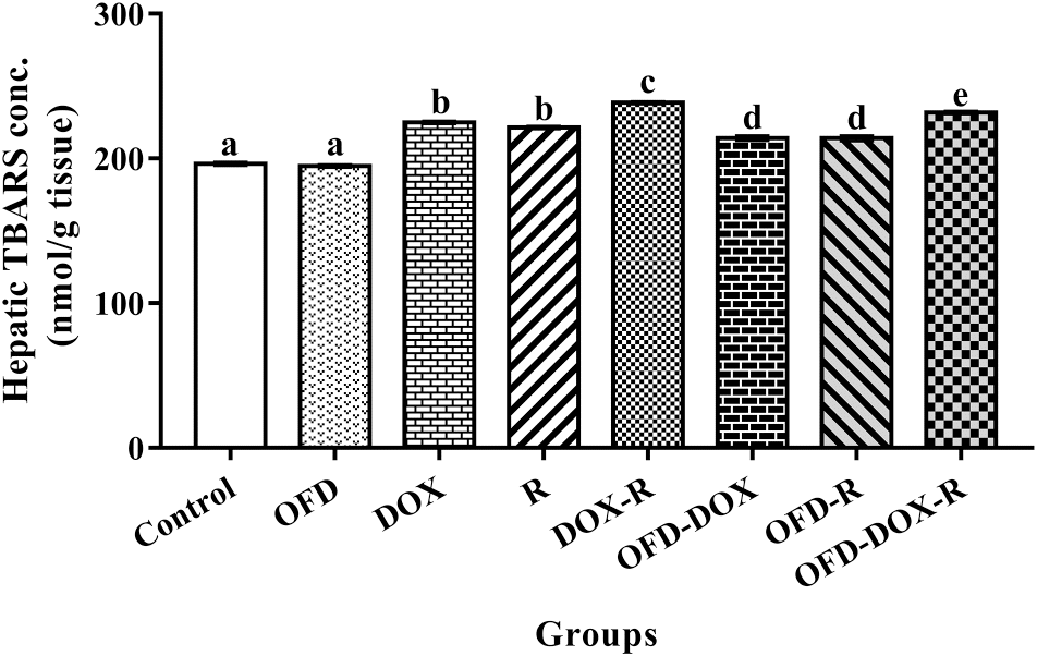
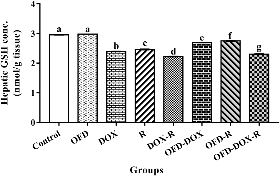
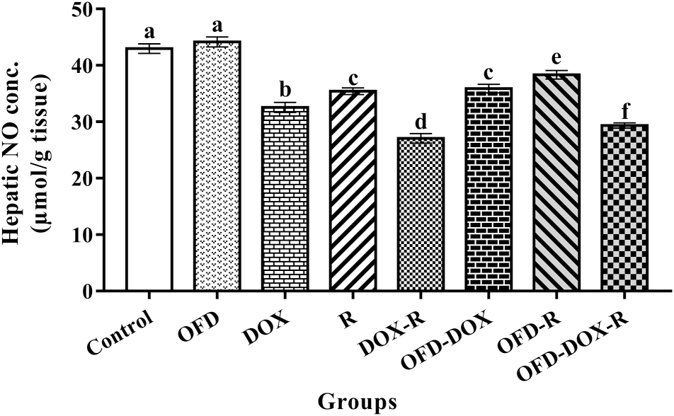
The DOX-treated and (or) R-treated rats showed a significant increase (p < 0.05) in hepatic TBARS and a significant decrease (p < 0.05) in hepatic GSH and NO concentrations compared with the corresponding values in the control group (Figs. 1, 2, and 3).
Fig. 1.
Fig. 1. The protective effects of olive oil with fig and date palm fruit extracts (OFD) on hepatic thiobarbituric acid reactive substances (TBARS) concentration in rats treated with doxorubicin (DOX) and (or) γ-radiation (R). Columns not sharing letters in common are significant with each other at p < 0.05.
Fig. 2.
Fig. 2. The protective effects of olive oil with fig and date palm fruit extracts (OFD) on hepatic reduced glutathione (GSH) level in rats treated with doxorubicin (DOX) and (or) γ-radiation (R). Columns not sharing letters in common are significant with each other at p < 0.05.
Fig. 3.
Fig. 3. The protective effects of olive oil with fig and date palm fruit extracts (OFD) on hepatic nitric oxide (NO) level in rats treated with doxorubicin (DOX) and (or) γ-radiation (R). Columns not sharing letters in common are significant with each other at p < 0.05.
In addition, the DOX-treated and (or) R-treated groups showed a significant increase (p < 0.05) in the serum levels of TG, TC, LDL-C, TG/HDL-C, TC/HDL-C, and LDL-C/HDL-C risk ratios, and a significant decrease (p < 0.05) in serum HDL-C compared with the corresponding values in the control group (Table 1).
Table 1.
Table 1. The protective effects of olive oil with fig and date palm fruit extracts on the serum lipid profile and lipid risk ratios of rats treated with doxorubicin and (or) γ-radiation.
Treatment groupTG (mg/dL)TC (mg/dL)LDL-C (mg/dL)HDL-C (mg/dL)TG/HDL-C risk ratio (mg/dL)TC/HDL-C risk ratio (mg/dL)LDL-C/HDL-C risk ratio (mg/dL)
Control80.21 ± 1.15a98.62 ± 0.95a17.48 ± 1.51a65.09 ± 1.62a1.23 ± 0.03a1.52 ± 0.03a0.27 ± 0.02a
OFD81.64 ± 1.62a98.09 ± 2.04a16.27 ± 1.94a65.49 ± 2.05a1.25 ± 0.05a1.50 ± 0.03a0.25 ± 0.03a
DOX129.42 ± 2.09b130.57 ± 2.49b59.41 ± 2.66b45.27 ± 1.32b2.87 ± 0.10b2.89 ± 0.10b1.32 ± 0.08b
R112.78 ± 2.15c127.80 ± 1.67bc56.34 ± 2.34b48.90 ± 1.26c2.31 ± 0.07c2.62 ± 0.07c1.16 ± 0.07c
DOX-R139.88 ± 2.08d144.47 ± 1.45d80.06 ± 1.66c36.42 ± 1.15d3.85 ± 0.09d3.98 ± 0.12d2.21 ± 0.10d
OFD-DOX97.45 ± 1.37e115.78 ± 1.19e45.68 ± 2.17d50.61 ± 2.05c1.94 ± 0.08e2.30 ± 0.09e0.91 ± 0.08e
OFD-R93.02 ± 2.19f111.72 ± 2.12f38.37 ± 2.31e54.74 ± 1.60e1.70 ± 0.06f2.04 ± 0.06f0.70 ± 0.05f
OFD-DOX-R114.74 ± 2.23c124.82 ± 1.87c59.86 ± 2.31b42.00 ± 1.30b2.74 ± 0.08b2.98 ± 0.10b1.43 ± 0.09b

Note: Results are expressed as mean ± SE. For each parameter, values not sharing common letters are significant with each other at p < 0.05; OFD, olive oil with fig and date palm fruit extracts; DOX, doxorubicin; R, irradiation; TG, triglycerides; TC, total cholesterol; LDL-C, low-density lipoprotein cholesterol; HDL-C, high-density lipoprotein cholesterol.

Moreover, the DOX-treated and (or) R-treated groups showed a significant decrease (p < 0.05) in RBC count, Hb concentration, Hct percentage, platelet count, WBC count, and lymphocyte percentage, and a significant increase (p < 0.05) in neutrophil and monocyte percentage compared with the corresponding values in the control group (Table 2).
Table 2.
Table 2. The protective effects of olive oil with fig and date palm fruit extracts on hematological parameters in rats treated with doxorubicin and (or) γ-radiation.
      Differential leucocyte count
Treatment groupRBC count × 106/mm3)Hb concentration (g/dL)Hct (%)Platelet count (103/mm3)WBC count (103/mm3)Lymphocytes (%)Neutrophils (%)Monocytes (%)
Control8.57 ± 0.06a15.62 ± 0.09a47.02 ± 0.17a936.7 ± 2.79a9.71 ± 0.05a72.2 ± 0.19a19.5 ± 0.16a4 ± 0.21a
OFD8.75 ± 0.08a15.90 ± 0.14a47.75 ± 0.34a937.1 ± 3.73a9.80 ± 0.06a71.6 ± 0.28a19.5 ± 0.21a4.2 ± 0.37a
DOX6.49 ± 0.06b11.85 ± 0.13b35.62 ± 0.37b702 ± 2.87b6.81 ± 0.03b65.5 ± 0.21b22.4 ± 0.28b6.4 ± 0.21b
R6.81 ± 0.04c12.44 ± 0.08c37.25 ± 0.39c714.2 ± 2.38c7.37 ± 0.05c67 ± 0.27c21.6 ± 0.21c6.4 ± 0.34b
DOX-R6.05 ± 0.06d11.03 ± 0.10d33.19 ± 0.31d664.9 ± 3.19d5.89 ± 0.03d62.8 ± 0.25d23.2 ± 0.25d6.9 ± 0.35b
OFD-DOX7.49 ± 0.12e13.66 ± 0.14e41.28 ± 0.47e728.8 ± 3.48e7.48 ± 0.03e68.7 ± 0.33e20.9 ± 0.30e5.4 ± 0.34c
OFD-R7.76 ± 0.08f13.95 ± 0.18e42.02 ± 0.56e745.5 ± 3.66f7.78 ± 0.03f68.7 ± 0.38e20.4 ± 0.21e6.2 ± 0.50bc
OFD-DOX-R7.14 ± 0.11g12.90 ± 0.16f38.62 ± 0.43f679.8 ± 3.00g6.35 ± 0.05g63.8 ± 0.25f22.7 ± 0.19bd6.9 ± 0.44b

Note: Results are expressed as mean ± SE. For each parameter, values not sharing common letters are significant with each other at p < 0.05. OFD, olive oil with fig and date palm extracts; DOX, doxorubicin; R, irradiation; RBC, red blood cell; Hb, hemoglobin; Hct, hematocrit; WBC, white blood cell.

The pretreatment of the DOX and (or) R treatment groups with OFD for two weeks as well as OFD treatment during the four weeks of the experiment significantly reduced (p < 0.05) the hepatic TBARS and significantly increased (p < 0.05) the hepatic GSH and NO compared with the DOX and (or) R treatment groups (Figs. 1, 2, and 3).
In addition, pretreatment of the DOX and (or) R treatment groups with OFD for two weeks as well as OFD treatment during the four weeks of the experiment significantly reduced (p < 0.05) the serum TG, TC, LDL-C, TG/HDL-C, TC/HDL-C, and LDL-C/HDL-C risk ratios and significantly increased (p < 0.05) serum HDL-C compared with the DOX and (or) R treatment groups (Table 1).
Moreover, pretreatment of the DOX and (or) R treatment groups with OFD for two weeks as well as OFD treatment during the four weeks of the experiment significantly increased (p < 0.05) RBC count, Hb concentration, Hct percentage, platelet count, WBC count, and lymphocyte percentage and significantly decreased (p < 0.05) neutrophil and monocyte percentages compared with the DOX and (or) R treatment groups (Table 2).

Discussion

Oxidative stress and generation of ROS may contribute to DOX and (or) R cytotoxicity during chemotherapy and radiotherapy (Elsadek et al. 2017; Fathy et al. 2017b). Among the major forms of cellular damage induced by DOX and (or) R exposure are DNA damage and lipid peroxidation. The increased levels of hepatic TBARS in the DOX and (or) R treatment groups and the decreased levels of GSH and NO compared with the control group indicate high levels of oxidative stress.
NO is a small, diffusible, highly reactive molecule that can generate oxidative stress (Millar 2004; Fathy et al. 2018). The significant decrease in NO levels recorded in the liver tissue after DOX and (or) R treatment might be the result of its interaction with superoxide to form peroxynitrite (Pryor and Squadrito 1995).
GSH deficiency contributes to oxidative stress and, therefore, may play a key role in aging and many diseases (Wu et al. 2004). In addition, GSH depletion after DOX and (or) R exposure may result from its diffusion through impaired cellular membranes and (or) inhibition of GSH synthetase and glutathione reductase enzymes (Zahran et al. 2006). Moreover, Srinivasan et al. (2006) demonstrated that decreased levels of GSH from oxidative stress might be due to its utilization by ROS.
The products of lipid peroxidation (TBARS) are used as an indicator of tissue damage (Zhou et al. 2006; Fathy et al. 2017a). The observed increase in TBARS levels may be attributed to increased ROS in the aqueous media of the cells and the interaction of the hydroxyl radical with the polyunsaturated fatty acids of membranes in the phospholipid portion of the cellular membranes initiating lipid peroxidation and the resulting damage of the cell membranes (Azab et al. 2001).
The depletion of NO and GSH and the increase in TBARS in DOX-treated and (or) R-treated groups match the results of studies by Bhatia and Jain (2004) and Abd Elbaky et al. (2010) who reported a significant depletion in the antioxidant system accompanied by the increase in lipid peroxides in rats treated with DOX or R.
Our results also showed an increase in the lipid profile and lipid risk ratios in the serum of rats treated with DOX and (or) R. Hypercholesterolemia conditions might result from the stimulation of cholesterol synthesis in the liver due to its release from tissues, the destruction of cell membranes and an increase in the rate of cholesterol biosynthesis in the liver and other tissues (Fathy 2014; Al-Saedi et al. 2015), the mobilization of fats from the adipose tissues into the bloodstream and mitochondrial dysfunction (Said and Azab 2006), interesterification, or random reaction changes in fatty acid positional distribution and the solid fat content of fats, which may consequently affect fat absorption and metabolism (Wang et al. 2016). Moreover, (Bok et al. 1999) attributed hypercholesterolemia to the increased activation of 3-Hydroxyl-3-methyl glutaryl coenzyme A (HMG-CoA) reductase enzyme, the key regulatory enzyme in the reduction of the overall process of cholesterol synthesis. Molchanova and Ahlers (1989) attributed the increase in serum triglyceride levels to the inhibition of lipoprotein lipase activity as well as an increase in cell damage and an efflux of triglycerides from the adipose tissues. Free radicals impair liver functions and cause hormonal imbalance, which induces hyperlipidemia through its multiple effects on lipid metabolism, including increased synthesis of cholesterol, triglyceride, and LDL-C (Bowden et al. 1989; Fathy et al. 2017a).
The hematopoietic system is highly sensitive to DOX and ionizing radiation. The reduction in the hematological parameters from DOX and (or) ionizing radiation treatments might be due to damage in the hematopoietic system (Fathy 2014) or the increased permeability of cell membrane, which in turn caused osmotic swelling and erythrocyte hemolysis (Asmis et al. 2005). It might also be due to the increased destruction of mature cells or increased plasma volume (Patil et al. 2008), or a decreased hemoglobin affinity for oxygen that induced hypoxia via diminished O2 transport from the lungs to the blood and decreased O2 release from oxyhemoglobin to the tissues (Jagetia et al. 2006).
OFD is rich in polyphenolic substances, which have received widespread attention because of their potential for preventing some common chronic diseases. Polyphenols are reported to have anti-inflammatory, antioxidant, antidiabetic, and hepatoprotective effects (Bashandy et al. 2014; El Arem et al. 2014).
OFD treatment for two weeks prior to DOX and (or) R treatment and during the four weeks of treatment during the experiment protected against oxidative stress. This was demonstrated by significantly ameliorated oxidative stress markers (TBARS, GSH, and NO) in the liver tissue compared with the DOX-treated and (or) R-treated rats. This might be a result of the protective action of OFD active ingredients modifying membrane organization and their ability to scavenge oxidation-initiating agents (Bashandy et al. 2014; Fathy et al. 2018). The antioxidant effect of OFD is mainly due to phenolic compounds, which are able to donate a hydrogen atom to the free radicals, thus stopping the chain reaction propagation during the lipid peroxidation process (Sanchez-Mareno et al. 1998). The two-week pretreatment of DOX-treated and (or) R-treated rats with OFD was also found to increase GSH and NO production. The antioxidant properties of GSH and NO stem from their reaction with oxygen, carbon, and nitrogen-centered radicals, and they play a scavenger role against free radical attack (Grisham et al. 1999). In addition, NO modulates the inflammatory response by inhibiting the formation of proinflammatory lipids (Rubbo et al. 1994).
In vitro experiments have shown that olive oil protects LDL-C from lipid peroxidation (Owen et al. 2000). Mounting evidence indicates that OFD is remarkably rich in effective phenolic antioxidants that could provide protection by free radical scavenging and inhibiting oxidative damage (Rubió et al. 2014; Bashandy et al. 2016; Alfieri et al. 2017).
The results of our study are in agreement with the findings of Gorinstein et al. (2002) who reported that polyphenols decreased plasma LDL-C levels and prevented their oxidation in vivo. The mechanism of this hypocholesterolemic action may be the inhibition of dietary cholesterol absorption in the intestine, inhibition of cholesterol production by the liver (Krzeminski et al. 2003), or stimulation of the biliary secretion and cholesterol excretion in the feces (Prasad and Kalra 1993; Katan et al. 1995). The intake of unsaturated fat decreases plasma cholesterol, whereas the intake of saturated fat increases it (Grundy and Denke 1990; Visioli et al. 2005; Alfieri et al. 2017). Moreover, Fathy (2014) found that the administration of fig extract and (or) olive oil to R-treated rats significantly ameliorated the lipid profile parameters and lipid risk ratios.
The administration of OFD to DOX-treated and (or) R-treated rats improves the hematological parameters (RBC, Hb, Hct, platelets, total, and differential WBCs), which might be attributed to increased antioxidant enzymes leading to diminished oxidative stress in bone marrow and the spleen. The results of our study are in agreement with the results of studies by Viola and Viola (2009) and Fathy et al. (2018) who attributed the improved hematological parameters to oleuropein, a component of olive oil, which also exerts a favorable action on the platelets.

Conclusion

Our study demonstrated that the administration of OFD to rats treated with DOX and (or) R ameliorated the oxidative stress markers in the liver tissue and improved the hematological parameters as well as the lipid profile and lipid risk ratios. These novel findings revealed the synergistic effect of the OFD combination to produce a broad spectrum of antioxidative activities that effectively defend against free radical attack and combat oxidative stress.

Acknowledgements

The authors are very grateful for Experiments and Advanced Pharmaceutical Research Unit (EAPRU) and Center for Drug Discovery and Development Research at the Faculty of Pharmacy, Ain Shams University, Abassia, Cairo 11566, Egypt for kind help in performing the extract procedures and analyses.

References

Abd Elbaky NA, Ali AA, and Ahmed RA. 2010. Cardioprotective effect of simvastatin on doxorubicin induced oxidative cardiotoxicity in rats. Journal of Basic and Applied Sciences, 6(1): 29–38.
Al-Qarawi AA, Abdel-Rahman H, Ali BH, Mousa HM, and El-Mougy SA. 2005. The ameliorative effect of dates (Phoenix dactylifera L.) on ethanol-induced gastric ulcer in rats. Journal of Ethnopharmacology, 98(3): 313–317.
Al-Saedi HF, Al-Zubaidy AA, Khattab YI, and Sahib HB. 2015. Effect of pentoxifylline against doxorubicin-induced nephrotoxicity in rabbits. International Journal of Pharmaceutical Sciences Review and Research, 30(1): 195–199 [online]: Available from globalresearchonline.net/journalcontents/v30-1/36.pdf.
Alfieri A, Imperlini E, Nigro E, Vitucci D, Orrù S, Daniele A, et al. 2017. Effects of plant oil interesterified triacylglycerols on lipemia and human health. International Journal of Molecular Sciences, 19(1): 104.
Allain CC, Poon LS, Chan CS, Richmond WG, and Fu PC. 1974. Enzymatic determination of total serum cholesterol. Clinical Chemistry, 20(4): 470–475.
Asmis R, Wang Y, Xu L, Kisgati M, Begley JG, and Mieyal JJ. 2005. A novel thiol oxidation-based mechanism for adria-mycin-induced cell injury in human macrophages. FASEB Journal, 19: 1866–1868.
Azab KS, Saada HN, and Said UZ. 2001. The action of long-term treatment with Coenzyme Q10 in minimizing radiation induced damage. Arab Journal of Nuclear Sciences and Applications, 34(1): 283–289 [online]: Available from inis.iaea.org/search/searchsinglerecord.aspx?recordsFor=SingleRecord&RN=33018612.
Baatout S, Jacquet P, Derradji H, Ooms D, Michaux A, and Mergeay M. 2004. Study of the combined effect of X-irradiation and epigallocatechin-gallate (a tea component) on the growth inhibition and induction of apoptosis in human cancer cell lines. Oncology Reports, 12(1): 159–167.
Bashandy MA, Abd El-Rasheid HG, Hasan HF, and Fathy AH. 2014. Protective and therapeutic effects of olive oil and Ficus carica as natural antioxidants on some biochemical parameters in liver of γ-irradiated male albino rats. Al Azhar Bulletin of Science, 25(1): 1–16.
Bashandy MA, Abd-el-aal A, Ibrahim DF, and El-sharkawy MA. 2016. Protective effects of date palm extract as natural antioxidants on hepatotoxicity induced by Cerastes cerastes venom in albino rats. International Journal of Advanced Research, 4: 1189–1195.
Beutler E, Duron O, and Kelly BM. 1963. Improved method for the determination of blood glutathione. The Journal of Laboratory and Clinical Medicine, 61(5): 882–888.
Bhatia AL, and Jain M. 2004. Spinacia olleracea L. protects against gamma radiations: a study on glutathione and lipid peroxidation in mouse liver. Phytomedicine, 11(7–8): 607–615.
Bok SH, Lee SH, Park YB, Bae KH, Son KH, Jeong TS, et al. 1999. Plasma and hepatic cholesterol and hepatic activities of 3-hydroxy-3-methyl-glutaryl-CoA reductase and acyl CoA: cholesterol transferase are lower in rats fed citrus peel extract or a mixture of citrus bioflavonoids. Journal of Nutrition, 129(6): 1182–1185.
Bowden DA, McLean P, Steinmetz A, Fontana D, Matthys C, Warnick GR, et al. 1989. Lipoprotein, apolipoprotein, and lipolytic enzyme changes following estrogen administration in postmenopausal women. The Journal of Lipid Research, 30: 1895–1906. Available from jlr.org/content/30/12/1895.full.pdf+html.
Burstein M, Scholnick HR, and Morfin R. 1970. Rapid method for the isolation of lipoproteins from human serum by precipitation with polyanions. The Journal of Lipid Research, 11: 583–595. Available from jlr.org/content/11/6/583.full.pdf+html.
El Arem A, Saafi EB, Ghrairi F, Thouri A, Zekri M, Ayed A, et al. 2014. Aqueous date fruit extract protects against lipid peroxidation and improves antioxidant status in the liver of rats subchronically exposed to trichloroacetic acid. Journal of Physiology and Biochemistry, 70(2): 451–464.
Elsadek B, Mansour A, Saleem T, Warnecke A, and Kratz F. 2017. The antitumor activity of a lactosaminated albumin conjugate of doxorubicin in a chemically induced hepatocellular carcinoma rat model compared to sorafenib. Digestive and Liver Disease, 49(2): 213–222.
Fathy AH. 2014. Influence of extracted Ficus and olive oil on some physiological and biochemical parameters in gamma irradiated albino rats. In Zoology. Al-Azhar University, Nasr City, Cairo, Egypt. 285 p.
Fathy AH, Bashandy MA, Bashandy SAE, Mansour AM, and Elsadek B. 2017a. Sequential analysis and staging of a diethylnitrosamine-induced hepatocellular carcinoma in male Wistar albino rat model. Canadian Journal of Physiology and Pharmacology, 95(12): 1462–1472.
Fathy AH, Bashandy MA, Mansour AM, Azab KS, and Bashandy SAE. 2017b. Hepatoprotective effects of olive oil with fig and date-palm fruit extracts in rats treated with doxorubicin and gamma radiation. Advances in Pharmaceutical and Ethnomedicines, 5(1): 8–15.
Fathy AH, Bashandy MA, Bashandy SA, and Mansour AM. 2018. The beneficial effect of natural antioxidants from olive oil with fig and date palm fruit extracts on biochemical and hematological parameters in rats during diethylnitrosamine-induced carcinogenesis. FACETS, 3: 584–597.
Fossati P, and Prencipe L. 1982. Serum triglycerides determined colorimetrically with an enzyme that produces hydrogen peroxide. Clinical Chemistry, 28(10): 2077–2080. Available from clinchem.aaccjnls.org/content/28/10/2077.
Gilani AH, Mehmood MH, Janbaz KH, Khan AU, and Saeed SA. 2008. Ethnopharmacological studies on antispasmodic and antiplatelet activities of Ficus carica. Journal of Ethnopharmacology, 119(1): 1–5.
Gorinstein S, Leontowicz H, Lojek A, Leontowicz M, Ciz M, Krzeminski R, et al. 2002. Olive oils improve lipid metabolism and increase antioxidant potential in rats fed diets containing cholesterol. Journal of Agricultural and Food Chemistry, 50(21): 6102–6108.
Grisham MB, Jourd’Heuil D, and Wink DA. 1999. Nitric oxide. I. Physiological chemistry of nitric oxide and its metabolites: implications in inflammation. American Journal of Physiology, 276(2 Pt 1): G315–321. Available from ajpgi.physiology.org/content/ajpgi/276/2/G315.full.pdf.
Grundy SM, and Denke MA. 1990. Dietary influences on serum lipids and lipoproteins. Journal of Lipid Research, 31(7): 1149–1172. Available from jlr.org/content/31/7/1149.full.pdf.
Institute of Laboratory Animal Resources. 1996. Guide for the care and use of laboratory animals. 8th edition. Committee for the update of the guide and use of laboratory animals. National Research Council of the National Academies, National Academy Press, Washington, D.C.
Jagetia GC, Venkatesh P, Archana P, Krishnanand BR, and Baliga MS. 2006. Effects of Aegle marmelos (L.) Correa on the peripheral blood and small intestine of mice exposed to gamma radiation. Journal of Environmental Pathology, Toxicology and Oncology, 25(4): 611–624.
Kamiloglu S, Pasli AA, Ozcelik B, and Capanoglu E. 2014. Evaluating the in vitro bioaccessibility of phenolics and antioxidant activity during consumption of dried fruits with nuts. LWT-Food Science and Technology, 56(2): 284–289.
Karbowink M, and Reiter RJ. 2000. Antioxidative effects of melatonin in protection against cellular damage caused by ionizing radiation. Proceedings of the Society for Experimental Biology and Medicine, 225(1): 9–22.
Katan MB, Zock PL, and Mensink RP. 1995. Dietary oils, serum lipoproteins, and coronary heart disease. The American Journal of Clinical Nutrition, 61(Suppl. 6): 1368S–1373S.
Krzeminski R, Gorinstein S, Leontowicz H, Leontowicz M, Gralak M, Czerwinski J, et al. 2003. Effect of different olive oils on bile excretion in rats fed cholesterol-containing and cholesterol-free diets. Journal of Agricultural and Food Chemistry, 51(19): 5774–5779.
Millar TM. 2004. Peroxynitrite formation from the simultaneous reduction of nitrite and oxygen by xanthine oxidase. FEBS Letters, 562(1–3): 129–133.
Molchanova A, and Ahlers I. 1989. Changes in the cellularity and lipid concentration of the bone marrow in rats after a single gamma-ray irradiation. Radiobiologiia, 29(2): 271–274.
Montgomery HA, and Dymock JF. 1961. The determination of nitrite in water. Analyst, 86: 414–416.
Owen RW, Mier W, Giacosa A, Hull WE, Spiegelhalder B, and Bartsch H. 2000. Phenolic compounds and squalene in olive oils: the concentration and antioxidant potential of total phenols, simple phenols, secoiridoids, lignansand squalene. Food and Chemical Toxicology, 38(8): 647–659.
Patil RR, Guhagarkar SA, and Devarajan PV. 2008. Engineered nanocarriers of doxorubicin: a current update. Critical Reviews in Therapeutic Drug Carrier Systems, 25(1): 1–61.
Prasad K, and Kalra J. 1993. Oxygen free radicals and hypercholesterolemic atherosclerosis: effect of vitamin E. American Heart Journal, 125(4): 958–973.
Pryor WA, and Squadrito GL. 1995. The chemistry of peroxynitrite: a product from the reaction of nitric oxide with superoxide. American Journal of Physiology, 268(5 Pt 1): L699–L722.
Reagan-Shaw S, Nihal M, and Ahmad N. 2008. Dose translation from animal to human studies revisited. FASEB Journal, 22(3): 659–661.
Rubbo H, Radi R, Trujillo M, Telleri R, Kalyanaraman B, Barnes S, et al. 1994. Nitric oxide regulation of superoxide and peroxynitrite-dependent lipid peroxidation. Formation of novel nitrogen-containing oxidized lipid derivatives. Journal of Biological Chemistry, 269: 26066–26075.
Rubió L, Macià A, Castell-Auví A, Pinent M, Blay MT, Ardévol A, et al. 2014. Effect of the co-occurring olive oil and thyme extracts on the phenolic bioaccessibility and bioavailability assessed by in vitro digestion and cell models. Food Chemistry, 149: 277–284.
Said UZ, and Azab KS. 2006. Efficacy of wheat germ oil in modulating radiation-induced heat damage in rats. Egyptian Journal of Radiation Sciences and Applications, 19(2): 433–452 [online]: Available from inis.iaea.org/search/searchsinglerecord.aspx?recordsFor=SingleRecord&RN=37071548.
Sanchez-Mareno C, Larrauri JA, and Saura-Calixto F. 1998. A procedure to measure the antiradical efficiency of polyphenols. Journal of the Science of Food and Agriculture, 76(2): 270–276.
Solomon A, Golubowicz S, Yablowicz Z, Grossman S, Bergman M, Gottlieb HE, et al. 2006. Antioxidant activities and anthocyanin content of fresh fruits of common fig (Ficus carica L.). Journal of Agricultural and Food Chemistry, 54(20): 7717–7723.
Srinivasan M, Sudheer AR, Pillai KR, Kumar PR, Sudhakaran PR, and Menon VP. 2006. Influence of ferulic acid on γ-radiation induced DNA damage, lipid peroxidation and antioxidant status in primary culture of isolated rat hepatocytes. Toxicology, 228(2–3): 249–258.
Turrens JF. 2003. Mitochondrial formation of reactive oxygen species. The Journal of Physiology, 552(2): 335–344.
Vayalil PK. 2002. Antioxidant and antimutagenic properties of aqueous extract of date fruit (Phoenix dactylifera L. Arecaceae). Journal of Agricultural and Food Chemistry, 50(3): 610–617.
Vinson JA, Zubik L, Bose P, Samman N, and Proch J. 2005. Dried fruits: excellent in vitro and in vivo antioxidants. Journal of the American College of Nutrition, 24(1): 44–50.
Viola P, and Viola M. 2009. Virgin olive oil as a fundamental nutritional component and skin protector. Clinics in Dermatology, 27(2): 159–165.
Visioli F, Caruso D, Grande S, Bosisio R, Villa M, Galli G, Sirtori C, and Galli C. 2005. Virgin Olive Oil Study (VOLOS): vasoprotective potential of extra virgin olive oil in mildly dyslipidemic patients. European Journal of Nutrition, 44(2): 121–127.
Wang T, Wang X, and Wang X. 2016. Effects of lipid structure changed by interesterification on melting property and lipemia. Lipids, 51(10): 1115–1126.
Wieland H, and Seidel D. 1982. Improved assessment of plasma lipoprotein patterns. IV. Simple preparation of a lyophilized control serum containing intact human plasma lipoproteins. Clinical Chemistry, 28(6): 1335–1337.
Wu G, Fang YZ, Yang S, Lupton JR, and Turner ND. 2004. Glutathione metabolism and its implications for health. The Journal of Nutrition, 134(3): 489–492.
Yoshioka T, Kawada K, Shimada T, and Mori M. 1979. Lipid peroxidation in maternal and cord blood and protective mechanism against activated-oxygen toxicity in the blood. American Journal of Obstetrics and Gynecology, 135(3): 372–376.
Zahran AM, Azab KS, and Abbady MI. 2006. Modulatory role of allopurinol on xanthine oxidoreductase system and antioxidant status in irradiated rats. Egyptian Journal of Radiation Sciences and Applications, 19(2): 373–388 [online]: Available from inis.iaea.org/search/search.aspx?orig_q=RN:37071546.
Zhou YH, Yu JP, Liu YF, Teng XJ, Ming M, Lv P, et al. 2006. Effects of Ginkgo biloba extract on inflammatory mediators (SOD, MDA, TNF-alpha, NF-kappaBp65, IL-6) in TNBS-induced colitis in rats. Mediators of Inflammation. 2006(5): 1–9.

Information & Authors

Information

Published In

cover image FACETS
FACETS
Volume 3Number 1October 2018
Pages: 722 - 735
Editor: Peter Zahradka

History

Received: 25 June 2017
Accepted: 7 May 2018
Version of record online: 9 July 2018

Data Availability Statement

All relevant data are within the paper.

Key Words

  1. olive oil
  2. fig fruit extract
  3. date palm fruit extract
  4. doxorubicin
  5. γ-radiation
  6. oxidative stress
  7. lipid profile
  8. hematological parameters

Sections

Subjects

Authors

Affiliations

Abdallah H. Fathy [email protected]
Department of Zoology, Faculty of Science, Al-Azhar University, Cairo, Egypt
Experiments and Advanced Pharmaceutical Research Unit, Faculty of Pharmacy, Ain Shams University, Cairo, Egypt
Mohamed A. Bashandy
Department of Zoology, Faculty of Science, Al-Azhar University, Cairo, Egypt
Samir A.E. Bashandy
Department of Pharmacology, Medical Division, National Research Center, Dokki, Giza, Egypt
Ahmed M. Mansour
Department of Pharmacology and Toxicology, Faculty of Pharmacy, Al-Azhar University, Cairo, Egypt
Khaled S. Azab
Department of Radiation Biology, National Center for Radiation Research and Technology, Atomic Energy Authority, Cairo, Egypt

Author Contributions

AHF, MAB, and AMM conceived and designed the study.
AHF and AMM performed the experiments/collected the data.
All analyzed and interpreted the data.
All contributed resources.
All drafted or revised the manuscript.

Competing Interests

The authors have declared that no competing interests exist.

Metrics & Citations

Metrics

Other Metrics

Citations

Cite As

Export Citations

If you have the appropriate software installed, you can download article citation data to the citation manager of your choice. Simply select your manager software from the list below and click Download.

Cited by

1. Therapeutic impact of purified Trichoderma viride L-asparaginase in murine model of liver cancer and in vitro Hep-G2 cell line
2. Antioxidant and Hepatoprotective Effects of Fig Fruit Extract with Olive Oil and Date-Palm Fruit Extract on Hepatic Toxicity of Oral Subchronic Exposure to Some Nanoparticles in Wistar Rats
3. Ameliorative Effects of some Natural Antioxidants against Blood and Cardiovascular Toxicity of Oral Subchronic Exposure to Silicon Dioxide, Aluminum Oxide, or Zinc Oxide Nanoparticles in Wistar Rats
4. Curcumin and vitamin C improve immunity of kidney via gene expression against diethyl nitrosamine induced nephrotoxicity in rats:In vivo and molecular docking studies
5. The protective effect of ethanol leaf extract of Annona muricata against doxorubicin toxicity via modulations of hematological, serum biochemical, antioxidant enzymes, and lipid peroxidation
6. Protective effect of fenofibrate against high‐fat–high‐fructose diet induced non‐obese NAFLD in rats
7. Modulatory effect of Ficus carica on oxidative stress and hematological changes induced by gamma-radiation in male albino rats

View Options

View options

PDF

View PDF

Get Access

Media

Media

Other

Tables

Share Options

Share

Share the article link

Share on social media