Open access

Distinct realized physiologies in green sea urchin (Strongylocentrotus droebachiensis) populations from barren and kelp habitats

Publication: FACETS
2 June 2022

Abstract

Overgrazing of habitat-forming kelps by sea urchins is reshaping reef seascapes in many temperate regions. Loss of kelp, in particular as a food source, may alter individual consumer physiology, which in turn may impair their ability to respond to climate warming. Here, we measured the temperature dependence of absolute and mass-independent oxygen consumption (M˙O2) using two different exposure protocols (acute exposure and temperature “ramping”), as proxies of realized physiology, between green sea urchin (Strongylocentrotus droebachiensis) populations from neighbouring barren and kelp habitats. Sea urchins from kelp habitats consumed 8%–78% more oxygen than sea urchins from barrens (across the range of temperatures tested (4–32 °C)) and had higher maximum M˙O2 values (by 26%). This was in part because kelp urchins typically had greater body masses. However, higher mass-independent M˙O2 values of kelp urchins suggest metabolic plasticity in response to habitat per se. In addition, the M˙O2 of sea urchins from kelp habitats was less sensitive to increases in temperature. We conclude that sea urchins from barren and kelp habitats of comparable body mass represent different energetic units. This highlights that habitat type can drive population-level variation that may shape urchins activities and environmental impact. Such variation should be integrated into energy-based models.

Introduction

Sea urchin populations are rapidly expanding across shallow rocky reef habitats in many temperate regions (Filbee-Dexter and Scheibling 2014). Urchins can reach high densities and form feeding (grazing) fronts that rapidly overgraze macroalgae, including bioengineering kelps (Filbee-Dexter and Scheibling 2014; Frey and Gagnon 2015; Ling et al. 2015). The proliferation of sea urchins, and the associated expansion of barrens, have been primarily linked to the over-exploitation of natural predators such as large groundfish and sea otters (Jessup et al. 2004; Pederson and Johnson 2006; Bonaviri et al. 2009; Sangil et al. 2012), but also to climate perturbations (e.g., heat waves) and disease-driven food web changes (McPherson et al. 2021; Smith et al. 2021). As kelp beds transition to barrens, habitat complexity, sediment accumulation and the availability of 3D-structures and shade decrease markedly, whereas wave exposure increases (Reed and Foster 1984; Rosman et al. 2007; Watanabe et al. 2016; Layton et al. 2019; Morris et al. 2020), and this limits both food sources and refugia. This shift to a more simplified ecosystem can impose strong “environmental filters”, defined as a set of environmental conditions that select a subset of species from a regional species’ pool (Lebrija-Trejos et al. 2010). Thus, the presence and absence of kelp may differentially select for subsets of traits or phenotypes (Kraft et al. 2015) and, hence, lead to shifts in assemblage structure as well as the functional and genetic structure of populations.
The paucity of food (including kelp) in sea urchin barrens suggests that there is a fundamental difference in the selection pressures on sea urchin phenotypes as compared to those in kelp habitats (Benjamin et al. 2010; Hollins et al. 2018; Duncan et al. 2019). Multiple species of macroalgae are commonly present in kelp habitats, and support preferential and selective feeding by grazers, whilst grazers in barrens often experience prolonged starvation and rely on drift kelp, filamentous algae, and biofilms for their nutrition (Vanderklift and Wernberg 2008; Filbee-Dexter and Scheibling 2014; Renaud et al. 2015). The quantity and quality of food influences metabolic rate, which ultimately controls the pace of life and underpins an organism’s physiology and functioning (Brown et al. 2004; Huey and Kingsolver 2019; Norin and Metcalfe 2019). Metabolic rate is the sum of all life-sustaining chemical reactions that create and use energy, and is typically estimated by measuring oxygen consumption (M˙O2). Metabolic plasticity is often observed under different environmental conditions and in relation to food (Norin and Metcalfe 2019). Therefore, metabolic rates may differ between sea urchins living in kelp and barren habitats because of contrasting food availabilities and quality.
Understanding how metabolic rates are shaped by a species’ habitat also has implications in the context of climate change and population-level resilience. Organisms that cannot meet their basic energetic needs because of resource limitations may not be able to regulate and optimize their metabolic response to environmental stress. Metabolic rate can be used as a proxy to estimate an organism’s overall physiological state and to characterize its sensitivity to environmental change (Silbiger et al. 2019), since metabolic rate fuels all organism functioning and is strongly temperature dependent (Boltzmann 1872; Gillooly et al. 2001; Dell et al. 2011). Populations with different physiological trait distributions can respond differently when exposed to challenging thermal conditions, such as heat waves (Padfield et al. 2016; Silbiger et al. 2019). For example, two populations with different thermal tolerance ranges, temperature optima (where performance is maximal), and thermal safety margins (i.e., the difference between a species’ optimal temperature and its critical upper thermal limit) may respond differently to the same heat wave event, with only one population experiencing adverse effects. Yet, there has been limited research into how habitat shifts may shape physiological trait distribution within coastal populations (but see: Bernhardt and Leslie 2013; Miller and Dowd 2019; Spindel et al. 2021) and what the implications are for climate resilience.
The northwest Atlantic represents a model system where green sea urchin (Strongylocentrotus droebachiensis (O.F. Müller, 1776)) populations typically reach high densities, and can transform kelp habitats into extensive barrens (Scheibling and Hatcher 2001). Green sea urchins in this region play a key ecological role as consumers, and exert strong top-down control on marine communities by removing foundational kelps (Scheibling et al. 1999; Gagnon et al. 2004; Scheibling and Lauzon-Guay 2007; Frey and Gagnon 2015). Even so, kelp beds and kelp patches of mainly Alaria esculenta and Laminaria spp. are present in the northwest Atlantic, in areas where sea urchin populations die off cyclically because of disease outbreaks or where high currents limit sea urchin grazing (Keats et al. 1990; Feehan and Scheibling 2014; Frey and Gagnon 2016). This system provides an opportunity to directly compare sea urchin populations in terms of their physiology from these adjacent, yet contrasting, habitats.
In the present study, we use a standardized experimental approach to, first, determine whether green sea urchins from barren and kelp habitats differ in their realized physiological state (metabolic rate and thermal sensitivity) by measuring their absolute, mass-independent, and mass-specific M˙O2 over a range of temperatures. Absolute M˙O2 estimates the energy required per unit time to maintain biological functions, whilst mass-specific M˙O2 gives metabolic rate scaled to the organisms’ mass (Peters 1983; Brown et al. 2004). Because absolute M˙O2 and body mass are typically correlated (Brown et al. 2004), and because sea urchin mass itself may vary across barrens and kelp habitats, we also calculate mass-independent M˙O2 to compare populations without the confounding effect of body mass. Second, we test whether the thermal sensitivity of metabolism differs between sea urchins from barren and kelp habitats. Third, we compare two experimental approaches to investigate the effects of short-term increases in sea water temperature on urchin metabolism: Assay Method 1 an “acute” temperature exposure protocol, where sea urchins are transferred to a novel (stable) temperature, and oxygen consumption is measured at that temperature; and Assay Method 2 a temperature “ramping” (dynamic) exposure protocol, in which the same set of individuals are exposed to stepwise increases in temperature (Terblanche et al. 2007). The latter experiment allowed us to construct thermal response curves (TRCs) for each individual (Huey and Stevenson 1979) and enabled us to evaluate an individual’s response to a temperature gradient. We used these two approaches to test if the different methods result in similar, or different, temperature-dependent changes in M˙O2 and to examine whether “heat-hardening” (i.e., an increase in heat tolerance following a sub-lethal exposure to elevated temperatures (Maness and Hutchison 1980)) occurs during temperature ramping.

Methods

Sea urchin collections

Green sea urchins (N = 225) were collected by snorkelers from three sites along the northeastern arm of the Avalon Peninsula: Biscayan Cove, Tors Cove, and Bauline (ordered by collection date; see Fig. 1 and Table 1 for details on the collection sites). Sites were chosen as comparable replicates of barren and kelp habitats with similar depth profiles, extent and exposure to waves, as well as shore access and distance from the laboratory. Sea urchins with test diameters of ∼7–8 cm were haphazardly hand collected from habitats within the same depth range (see Table 1, Supplementary Fig. S1) that resembled kelp or barrens habitats (i.e., with or without kelp) as assessed visually. Kelp areas were sections of rocky reef, at least 10 m × 5 m in size, with dense, continuous kelp cover, whilst barren areas (also at least 10 m × 5 m) were rocky reefs devoid of fleshy seaweeds, with bare rock substrate covered in encrusting coralline algae. Prior to collection, sea urchin densities were quantified in kelp and barren areas by counting the number of individuals in a 0.5 m × 0.5 m quadrat. Seven quadrats were haphazardly placed in each kelp and barren area of the three sites (N = 42 quadrats). Sea urchins collected in each habitat were kept separate and placed into individual seawater-filled coolers for immediate transport to the Ocean Science Centre (OSC) in Logy Bay, Newfoundland, within 2 h of collection. At the OSC, the sea urchins were placed into holding tanks with seawater at 14 °C (the average ambient summer temperature) and a 12-h light:12-h dark photoperiod and left to recover from transport and handling stress for 24 h. To measure the routine M˙O2 of the urchin populations in a “field-fresh” physiological state, M˙O2 measurements were started after the initial 24-h recovery period and completed within seven days of collection. Sea urchins were not fed at any point to avoid post-feeding increases in metabolic rate (i.e., specific dynamic action).
Fig. 1.
Fig. 1. The map shows green sea urchin collection sites along the easternmost side of the Avalon Peninsula, Newfoundland (NL), Canada (plot inset shows the province of NL, with the red box outlining the Avalon Peninsula). Urchins were hand collected from Biscayan Cove, Tors Cove, and Bauline (see Table 1 for further details). Panels on the right (A-D) show kelp beds (A, B) and sea urchin barrens (C, D) on the Avalon Peninsula. Pictures were taken by snorkelers at ∼4 m depth in August 2020. Panels B and C show count quadrats with a size of 0.5 m × 0.5 m, placed in a kelp bed and barren, respectively.
Table 1.
Table 1. Details on the green sea urchin collection sites used in these experiments.
Collection siteLatitude (°)Longitude (°)Collection depth (m)Date (DD-MM-YY)SW Temp.ProtocolNo. of sea urchins collected
Biscayan Cove47.80394752.7870871.4–5.612-08-2014 °CAcute72
Tors Cove47.21230252.8449152.5–4.506-09-2011 °CAcute72
Bauline47.72245652.8350110.7–3.021-09-2011 °CAcute72
Biscayan Cove47.80394752.7870872.0–3.006-11-207 °CRamping9

Note: The Protocol column indicates in which experiment a given group of urchins was used. Two experimental protocols were used; an acute temperature protocol and a ramping protocol (see Methods). The latter was used for the construction of individual thermal response curves. SW Temp. indicates ambient seawater temperature (°C) at the time of collection.

Experimental system for measuring oxygen consumption

We used a custom-built experimental system to measure the M˙O2 of individual sea urchins (see Supplementary Fig. S2). The system, built by the Technical Services Department at Memorial University of Newfoundland and Labrador, consists of a table with 10 removable acrylic chambers (each 650 mL in volume and 9 cm in diameter) with magnetic stir plates, located inside an insulated seawater tank (YETI Tundra cooler, 125 L; Austin, Texas, USA). A heater (submersible aquarium heater, 300 Watts; Aqueon, Franklin, Wisconsin, USA) and chiller (Isotemp Model 3016S; Fisher Scientific, Waltham, Massachusetts, USA) controlled by a thermostat (Inkbird ITC-308 Temperature Controller; Inkbird Tech, London, UK) were used to adjust seawater temperatures in the insulated cooler. A water pump connected to the chiller circulated the seawater from one end of the cooler to the other, ensuring consistent temperatures across the 10 chambers. Seawater within the cooler was continuously aerated with an air pump (Top Fin Aquarium, Air-200; PetSmart LLC, Phoenix, Arizona, USA) attached to an air stone. Each chamber was fitted with a temperature probe (PreSens dipping probe, Pt1000) and a fiber-optic oxygen probe (PreSens dipping probe, DP-PSt7-10-L2.5-ST10-YOP), and measurements of temperature and water oxygen level (in % saturation) were made every second via a computer running PreSens software (PreSens Measurement Studio 2, Version 3.0.3; Precision Sensing GmbH, Regensburg, Germany). Water oxygen level (in mL O2/L) was automatically calculated by the PreSens software. Oxygen probes were calibrated prior to each experimental set (sampling site) with air saturated, room temperature, seawater, and sodium sulfite (no O2; 1 g Na2SO3 dissolved in 100 mL of distilled water at room temperature).

Oxygen consumption measurements

Premeasurement procedures and containment of sea urchins

We used a standardized experimental approach to test for differences in the response of metabolism to temperature between sea urchin populations from barren and kelp habitats. To estimate routine metabolic rate at the eight seawater temperatures (4, 6, 10, 14, 18, 22, 26, or 30 °C), the M˙O2 of sea urchins was measured using closed respirometry with the system described above. We chose these temperatures to capture the entire response range (increase, peak, and decrease in oxygen consumption) of sea urchins to allow us to compare the response shape and limits across populations. The coldest and warmest temperatures were experimentally constrained by the heater and cooler capacities, and the intervals were chosen based on logistics and feasibility. Twelve hours before the start of each run, nine urchins were selected for measurement (four barren urchins and five kelp urchins or vice versa). These nine sea urchins were weighed (to the nearest 0.1 g), and had their individual volumetric displacement measured (volumetric displacement = volume of seawater with urchin – volume without urchin). One individual was placed into each of the nine chambers, with no urchin in the tenth chamber so that background oxygen consumption (i.e., due to microbial respiration) could be measured in a blank control chamber that contained only seawater. The chambers were then covered with fine-mesh netting to prevent the urchins from crawling out of the chamber and returned to the holding tank overnight to allow the urchins to adjust to confinement in the chambers and to recover from handling.

Acute temperature exposure protocol (Assay Method 1)

To examine the temperature-dependent M˙O2 response of sea urchins from all three sites to acute temperature changes, the YETI cooler was filled with fresh, filtered (10 μm) seawater and set to one of eight target temperatures: 4, 6, 10, 14, 18, 22, 26, or 30 °C. Each independent assay was conducted on a different day, and target temperatures were randomized for each day so that warmer temperatures did not relate to longer timespans during which the urchins were held in aquaria. For each assay, all 10 chambers (nine urchins, one blank) with mesh tops were moved from the holding tank to the insulated cooler (preset to the target temperature for the day), and the urchins were given 1 h at their new temperature (standardized to 1 h because this was the maximum time to equilibrium across all temperatures). Then M˙O2 measurements were made by closing the lids on each chamber and allowing oxygen levels in the chambers to fall by 5%–10%. This decrease in oxygen concentration allowed reliable estimates of M˙O2 to be obtained. The total measurement time varied from 15 min to ∼ 1.5 h, with shorter measurement times at higher temperatures because M˙O2 increases with temperature (Gillooly et al. 2001). Once the measurements were completed for each of the 10 chambers, all sea urchins included in the assay were removed from their chambers and immediately frozen prior to ash-free dry mass (AFDM) determination (see section Ash-free dry mass). The cooler and chambers were then emptied, cleaned with warm freshwater, and refilled with seawater for the next assay. The above procedures were repeated with new urchins until measurements were made at all target temperatures (N = 9 sea urchins per measurement; N = 72 sea urchins per collection site; N = 216 sea urchins in total across all sites and replicates).

Temperature ramping exposure protocol (Assay Method 2)

To examine the metabolic response of sea urchins to a temperature ramping protocol, and to assess how comparable values are between this method and the previous protocol (i.e., acute transfer to a new temperature), we constructed TRCs for individuals from Biscayan Cove (only one site was included because of logistical constraints). Nine sea urchins from a fresh collection were weighed and prepared as detailed in the “Premeasurement procedures” section. After an overnight recovery period, nine urchin-containing chambers and a blank chamber were placed into the cooler with temperature preset to 4 °C, and 1 h was allowed before testing began. Oxygen consumption measurements were then made at each of nine temperature steps: 4, 6, 10, 14, 18, 22, 26, 30, and 32 °C with a 30-min period between each temperature during which the next target temperature was reached and maintained (see Supplementary Fig. S3). The final temperature step (32 °C) was added to ensure we accurately defined the sea urchins’ TRC as completely as possible. The first experiment (acute protocol) showed that M˙O2 peaked at around 26 °C. The same nine sea urchins had their M˙O2 measured at all temperatures, and the chamber lids were removed and replaced with the mesh lids between measurements to allow for the replacement of seawater from the cooler in which the chambers were held. After the final measurement was taken, the sea urchins were immediately frozen.

Ash-free dry mass

We determined AFDM to quantify the amount of organic tissue per sea urchin, which corresponds to the amount of metabolically active tissue. To measure AFDM, empty aluminum weigh boats were first placed in a muffle furnace (500 °C) for 12 h to remove any trace of organic matter and, thereafter, stored in a sealed container until use. Sea urchins frozen at the end of both temperature challenges were thawed prior to weighing. The urchins were then placed on preweighed (to 0.001 g accuracy) weigh boats and dried in a combustion oven (at 60 °C) for 12–24 h until their dry mass stabilized. The sea urchins were then ashed in a muffle furnace at 500 °C for 12 h. AFDM (i.e., metabolically active tissue) was calculated as dry mass (with boat) minus ashed mass (with boat). Individual dry mass was calculated as dry mass minus the empty weigh-boat weight, where dry mass was the total mass of an individual (organic and inorganic) after drying. Finally, an individual’s inorganic mass was calculated as ashed mass minus the empty boat weight. This inorganic mass primarily represents an individual’s calcified endoskeleton (test), although small traces of sediment or inorganic gut content may also be present in the ashed sample.

Data processing and analyses

Values of M˙O2 were calculated for each individual using the respR package in R (Harianto et al. 2019). M˙O2 values were adjusted for salinity and water volume in the chamber (i.e., after correction for an individual’s volumetric displacement). Mass-independent M˙O2 was calculated by regressing absolute M˙O2 on individual wet body mass (nonlinear regression) and extracting the residuals. Mass-specific values were adjusted for wet mass or AFDM (i.e., absolute M˙O2 divided by mass), with the latter accounting for the mass of metabolically active tissue only. All M˙O2 measurements were also corrected for background respiration using the M˙O2 values for the blank chamber of each run. Background respiration values were typically < 0.1 mL O2/h. The M˙O2 data were visually inspected to confirm that a linear decrease in water % air saturation of 5%–10% occurred. We set a minimum r2 of 0.98 (Chabot et al. 2021) to identify nonlinear measurements and discarded eight measurements (all from Assay Method 1; Fig. S6) with an r2 value below this threshold.
To compare the temperature sensitivity of barren and kelp sea urchins, we calculated temperature coefficients (Q10 values) from the acute protocol assays, based on the mean absolute M˙O2 for each site and habitat group, and for the temperature ramping (TRC) protocol, using absolute values of M˙O2 for each individual sea urchin. Q10 values were calculated for each group or individual urchin based on the equation:
Q10=R2R1(10T2T1)
(1)
where R1 = M˙O2 at temperature T1, R2 = M˙O2 at temperature T2. Separate Q10 values were calculated for the lower temperature range (cold-range Q10; T1 = 4 °C and T2 = 14 °C) and the warm-range (i.e., temperatures above the average summer holding temperature (warm -range Q10; T1 = 14 °C and T2 = 26 °C)). We calculated these Q10 values separately because temperature sensitivity may change at more stressful temperatures that lie outside the sea urchins’ realized range. Additionally, we determined the maximum metabolic rate and estimated the temperature at which M˙O2 peaked (Tmax) from each thermal response curve by identifying the maximum M˙O2 achieved by each individual across temperatures and the temperature that corresponded with this maximum value. Finally, temperature-induced metabolic scope (AST) was calculated as the difference between the maximum recorded metabolic rate (MMRT; M˙O2 at 26 °C) and the lowest metabolic rate measured (LMRT; the M˙O2 at 4 °C).
To test for differences in the urchins’ M˙O2 between barren and kelp sites using Assay Method 1, we used generalized additive mixed models within the package “mgcv” in R (R Core Team 2014) using the “gamm” function (Wood 2011; Pinheiro et al. 2015). The random effect of site was included to account for variation in the response variables due to site. A random effect of “individual” was also used in models resulting from Assay Method 2 (the ramping protocol), since temperature ramping led to repeated measurements on the same individuals. Habitat (barrens vs. kelp) was included as a fixed effect in all models to test for habitat-dependent variation in the M˙O2 responses of the sea urchins, with temperature included as a covariate. For models with absolute M˙O2 as the response variable, we also included body mass as a covariate to account for the potentially confounding effect of individual mass.
We visually inspected Gaussian model fits to ensure test assumptions were met (normality of residuals and homogenous error structure), and we compared model results across different distribution families (Poisson and quasi-Poisson, both with a log-link function) to ensure the results were consistent. Additionally, we compared gamm results with results from linear mixed-model fits with a polynomial term (function “lme” in R package “nlme”), again ensuring reported model results were robust (Zuur et al. 2009).
To compare sea urchin mass (wet mass, ash-free dry mass, inorganic mass, and AFDM to wet mass ratio), metabolic parameters (MMRT and AST), and the thermal sensitivity of M˙O2 (i.e., Q10 values), we fit one-way or two-way analysis of variance (ANOVA) using the function “aov” in the R “stats” package (R Core Team 2014) with habitat, or habitat and site as main effects, and with an interaction term, as appropriate. We visually checked that test assumptions were met and ran a Shapiro–Wilk normality test using the function “shapiro.test” in the R stats package (R Core Team 2014). We performed Tukey’s HSD post-hoc tests to compare data between the three sites using the function “tukeyHSD” in the R stats package (R Core Team 2014).

Results

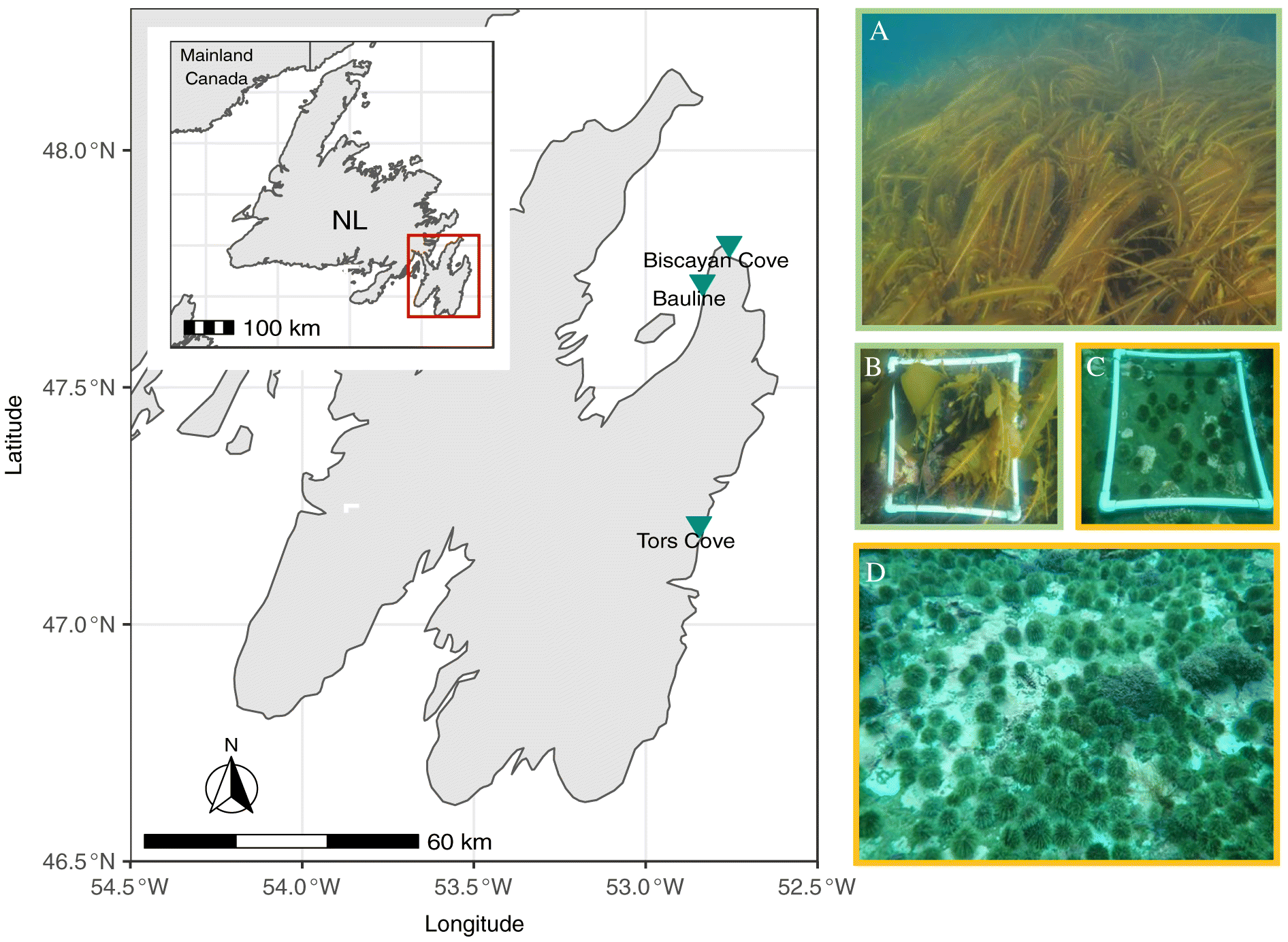
Sea urchin density averaged 25 ± 9 individuals per 0.25 m2 in barrens and 7 ± 5 individuals per 0.25 m2 in areas with kelp across the three sites (26 ± 7 vs. 6 ± 7 at Biscayan Cove, 22 ± 13 vs. 8 ± 4 at Tors Cove and 26 ± 9 vs. 7 ± 5 at Bauline, in kelp vs. barrens, respectively). Across the three sites, green sea urchins from kelp habitats had a greater overall mass (by 8%; two-way ANOVA: F(1,2) = 17.80, p < 0.01), more metabolically active tissue (i.e., higher AFDM values by 48% (two-way ANOVA: F(1,2) = 138.24, p < 0.01)) and more inorganic mass (by 16%; two-way ANOVA: F(1,2) = 23.66, p < 0.01) than sea urchins from barrens (Supplementary Fig. S4, Tables S5 and 
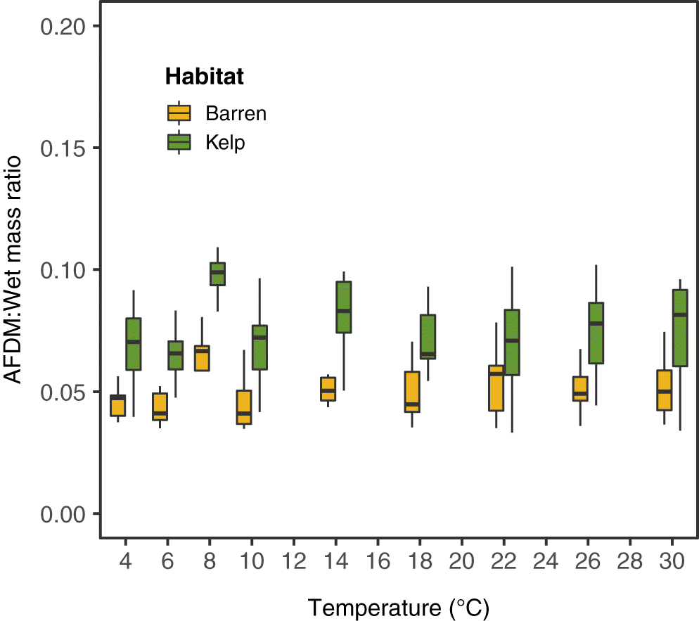
S6). Green sea urchin masses ranged across the sites from 23.3 g to 113.4 g (wet mass) and 0.9 g to 8.5 g (AFDM) in kelp and 15.2 g to 99.0 g (wet mass and) and 0.8 g to 5.3 g (AFDM) in barrens. Sea urchins from kelp habitats also had significantly higher AFDM to wet mass ratios (two-way ANOVA: F(1,2) = 47.15, p < 0.01; Fig. 2; Supplementary Table S5, Fig. S5) than individuals from barrens.
Fig. 2.
Fig. 2. Ash-free dry mass (AFDM) to wet mass ratios for green sea urchins collected from barren and kelp habitats in Newfoundland. The temperatures on the x-axis indicate the temperature steps each set of urchins was exposed to during oxygen consumption measurements. Data for each habitat and temperature combination represent 27 urchins, N = 216 in total. Boxplots show maximum, minimum and median values, as well as 25th and 75th percentile values.
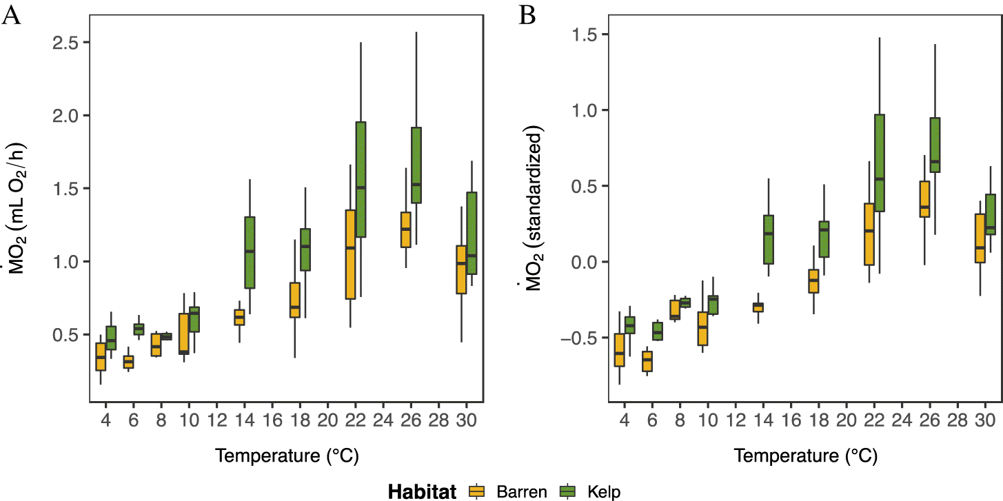
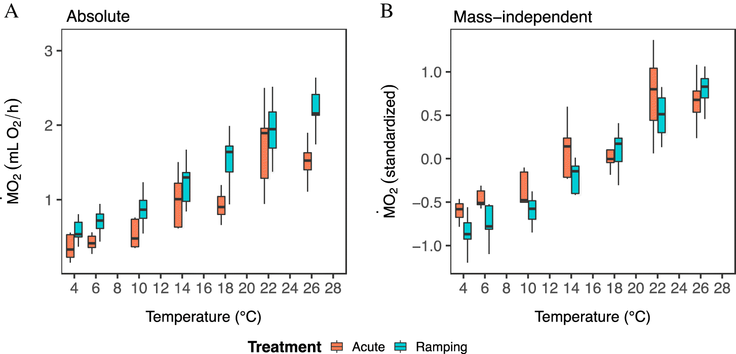
Green sea urchin populations from barren and kelp habitats differed in their oxygen consumption (Figs. 3 and 4). Overall, sea urchins from kelp habitats had significantly higher absolute M˙O2 values (by 8%–78%) than sea urchins from adjacent barrens (Figs. 3 and 4; Supplementary Table S1; Fig. S7) across the three study sites (Fig. 1), and this pattern was generally consistent across temperatures (Figs. 3 and 4). This difference was in part due to body mass (Table S1), as urchins from kelp habitats had a higher wet mass and a greater AFDM:wet-mass ratio (Fig. 2; Supplementary Figs. S4 and S5). However, M˙O2 per g of AFDM was, in fact, lower as compared to sea urchins from barren habitats (Supplementary Figs. S9 and S10). Further, after accounting for body mass, the mass-independent M˙O2 of sea urchins from kelp habitats was still significantly higher than that of sea urchins from barrens (Figs. 3A and 3B; Supplementary Table S1, Figs. S7C and S7D).
Fig. 3.
Fig. 3. Absolute (A) oxygen consumption (M˙O2) and mass-independent (B) oxygen consumption of green sea urchins collected from barren and kelp habitats as a function of experimental seawater temperature and measured using the acute protocol. Individuals were collected from three sites in Newfoundland and for each site, nine fresh urchins were acutely exposed to each temperature (N = 27 urchins per temperature). Boxplots show maximum, minimum, and median values as well as 25th and 75th percentile values. N = 216 urchins across all sites. Note: sea urchins from Bauline (N = 9 urchins) were mistakenly measured at 8 °C instead of 6 °C, thus boxplots at 6 °C represent sea urchins from Biscayan Cove and Tors Cove only (N = 18 urchins).
Fig. 4.
Fig. 4. Absolute oxygen consumption (M˙O2) of green sea urchins from barren and kelp habitats in Biscayan Cove when exposed to the ramping protocol. Boxplots (A) show maximum, minimum, and median absolute M˙O2 values as well as 25th and 75th percentile values. (B) Shows thermal response curves for each urchin (N = 9 urchins), with dashed lines and triangles denoting urchins from kelp habitats and solid lines and circles indicating those from urchin barrens.
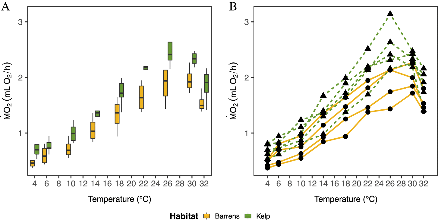
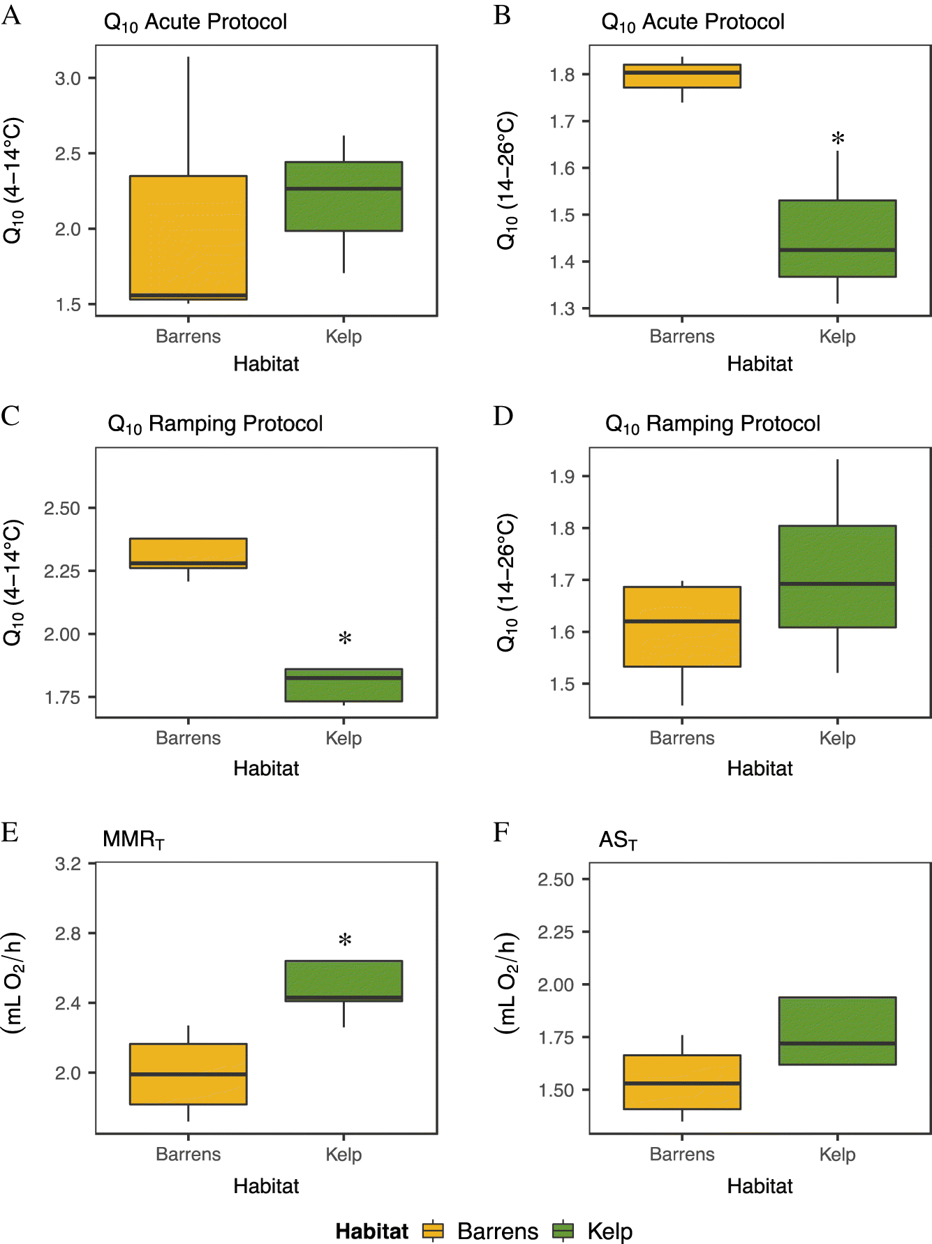
The temperature sensitivity (i.e., Q10 value) of sea urchin M˙O2 was significantly higher in animals from barrens than those from kelp habitats over the cold temperature range (Q10 4–14 °C; one-way ANOVA: F(1,6) = 27.47, p = 0.002, Supplementary Table S3), but only when assessed using the temperature ramping protocol (Table 2, Figs. 5A and 5C). Similarly, the temperature sensitivity of M˙O2 in sea urchins from barrens was significantly higher in the warm temperature range (Q10 14–26 °C) when measured using the acute protocol (one-way ANOVA: F(1,4) = 11.35, p = 0.028; Supplementary Table S3). Mean Q10 values ranged from 1.50 to 3.14, and from 1.31 to 1.84 across the two temperature ranges (4–14 °C and 14–26 °C, respectively) when sea urchins were exposed to the acute protocol. These values were similar to those measured using the ramping protocol, where Q10 values ranged from 1.72 to 2.69 and from 1.46 to 1.93, respectively.
Table 2.
Table 2. Temperature sensitivity (Q10 values) of absolute oxygen consumption, and parameters of metabolic performance, for green sea urchins collected from barren and kelp habitats at Bauline, Biscayan Cove and Tors Cove, Newfoundland.
ExperimentPopulation or replicateQ10 (4–14 °C)Q10 (14–26 °C)MMRT (mL O2/h)Tmax (°C)AST (mL O2/h)
AcuteBarrens Bauline1.561.801.3126NA
AcuteKelp Bauline1.811.641.4226NA
AcuteBarrens Biscayan Cove3.141.841.6326NA
AcuteKelp Biscayan Cove2.621.312.5022NA
AcuteBarrens Tors Cove1.501.741.8022NA
AcuteKelp Tors Cove2.261.422.5726NA
RampingBarrens #12.281.682.13261.63
RampingBarrens #22.671.452.27301.76
RampingBarrens #32.281.561.72301.35
RampingBarrens #42.211.701.85301.43
RampingKelp #11.861.802.64261.94
RampingKelp #21.831.932.26301.72
RampingKelp #31.731.522.43301.62
RampingKelp #41.721.612.41261.62
RampingKelp #52.69*1.693.14262.52

Note: Two separate Q10 values were calculated: i.e., 4–14 °C and 14–26 °C. The average water temperature during the summer was 14 °C. For the acute protocol, Q10 values were calculated based on the mean absolute M˙O2 values for each site/habitat combination, whereas Q10 values during the ramping protocol were calculated for individual urchins from Biscayan Cove. MMRT is the maximum absolute M˙O2 recorded. For the acute protocol, the individual with the highest absolute M˙O2 for each site and habitat is indicated. The temperature at which absolute M˙O2 was maximum is indicated by Tmax. AST is temperature-induced metabolic scope (MMRT – MR at 4 °C). One Q10 value in the cold range (4–14 °C) was excluded from the analysis after being confirmed as an outlier (marked by an * in the table) using a Grubb’s test (G = 1.852, U = 0.518, p = 0.171).

Fig. 5.
Fig. 5. Temperature sensitivity (Q10) of absolute oxygen consumption (M˙O2) in green sea urchins collected from barren and kelp habitats (A–D). Q10 values were calculated from 4–14 °C and 14–26 °C based on mean M˙O2 values for each site and habitat measured during the “acute” protocol (A and B, N = 3), and for each individual urchin using the “ramping” protocol (C and D, Biscayan Cove, see data in Fig. 4: N = 9). Note: mean summer water temperature was ∼14 °C. (E) The difference in maximum oxygen consumption (MMRT) between sea urchins from barren and kelp habitats. (F) The difference in temperature-induced metabolic scope (AST). Both parameters were calculated using data from the ramping protocol. N = 9 urchins per habitat type. Boxplots show maximum, minimum and median values, as well as 25th and 75th percentile values for each habitat type. Asterisks indicate a significant difference (* =  p < 0.05, ** = p < 0.01, ***p < 0.001) between values for sea urchins collected from barren and kelp habitats.
M˙O2 generally peaked between 26 and 30 °C when considering all site by habitat combinations (Table 2), with urchins inhabiting areas with kelp having significantly (by 26%) higher maximum M˙O2 values (one-way ANOVA; F(1,7) = 8.008, p = 0.025; Fig. 5E; Supplementary Table S3) than sea urchins from barrens. Despite the finding that sea urchins from kelp habitats had higher values for MMRT, there was no significant difference in the temperature-induced metabolic scope (AST, p = 0.254; Fig. 5F, Table 2; Supplementary Table S3). There were also no significant differences in values of MMRT or AST when standardized for wet mass (MMRT, p = 0.946; AST, p = 0.397) or AFDM (MMRT, p = 0.090; AST, p = 0.123), although sea urchins from kelp habitats had lower MMRT and AST values than sea urchins from barren habitats when standardized for AFDM (Supplementary Fig. S11). Overall, the M˙O2 of Biscayan Cove urchins did not differ significantly when measured using the two methods (acute temperature protocol vs. the ramping protocol: Fig. 6, Table S2, Fig. S8).
Fig. 6.
Fig. 6. Comparison of temperature-dependent changes in M˙O2 when measured using the acute (red) vs. the temperature ramping protocol (blue). (A) Absolute M˙O2 and (B) mass-independent M˙O2. Boxplots show maximum, minimum, and median values, as well as 25th and 75th percentile values, for each temperature exposure protocol (acute and ramping). N = 72 urchins across both protocols.

Discussion

We showed that green sea urchins from barren and kelp habitats in Newfoundland differ in their realized physiological states, with those from kelp habitats having higher metabolic rates, but reduced temperature sensitivity, compared to urchins from barren areas. The average urchin from areas with kelp consumed 8%–78% more oxygen than urchins from areas without kelp (i.e., barrens), across the range of typical ocean temperatures. The higher overall energetic requirements of sea urchins in kelp habitats were in part due to their greater mass. Yet, significant differences remained between sea urchins from areas with and without kelp when their mass-independent oxygen consumption rates were compared, which indicates that metabolic plasticity exists between habitat types regardless of mass effects. In contrast, when considering metabolism standardized per gram of animal, (i.e., AFDM) sea urchins from kelp habitats consumed less oxygen than barren urchins, which suggests a larger investment in energy-storing tissues with low oxygen demand in populations from areas with kelp. We conclude that sea urchin populations from kelp and barren habitats have fundamentally different mass-specific and mass-independent energy requirements and, hence, represent ecologically distinctive “units”. Such distinct populations may respond to, and interact with, their environment in unique but predictable ways.
Variation in individual energy requirements is ecologically important because biomass–energy relationships and the energetic status of a population form the basis of an organisms’ ability to respond to environmental variability. Energetic models, such as the metabolic theory of ecology (Brown et al. 2004) or dynamic energy budget models (Kooijman 1986), are built upon mass-energy scaling laws and are commonly used to predict organismal responses to environmental variability. Yet, most energetic models are average-based and predict a species’ mean response (rather than population specific responses) with unexplained, but often substantial, variation around the mean (Saito et al. 2021). This can lead to inaccurate predictions. Integrating habitat-based energy relationships into energetic models could explain some of this variation, and improve the forecasting of a species’ vulnerability to environmental change.
The differences we detected in the metabolic performance of green sea urchins from barrens and kelp habitats presumably relate to habitat characteristics and the physical challenges and environmental filters that each habitat presents (as summarized in the Introduction). Yet, differences in food availability or quality emerge amongst a number of abiotic and biotic differences as the most likely driver of differences in the energetic demand and physiology of sea urchins (Mueller and Diamond 2001; Huey and Kingsolver 2019). This is because populations in kelp habitats have access to nutritious and rich food sources, whilst high-density urchin barren populations experience prolonged periods of starvation and compete for scarce food sources such as drift kelp, encrusting algae, and biofilms (Norderhaug et al. 2003; Vanderklift and Wernberg 2008; Filbee-Dexter and Scheibling 2014; Renaud et al. 2015; Wells et al. 2017). Increased competition for scarce food resources in barrens intensifies food shortages, and this also appeared to be the case in the present study. Sea urchin densities were over three-fold higher in barren areas than in kelp areas across all sites.
Organisms that cannot meet their basic energetic demands because of reduced food availability or quality, or starvation, have limited capacity to regulate and optimize their metabolic response to environmental change (Boersma et al. 2008; O’Connor et al. 2009). Compared to sea urchins from kelp habitats, urchins from barrens in our experiments had lower absolute MMRT values (by 26%), and their M˙O2 was also more sensitive to increasing temperature (i.e., they had higher Q10 values). Perhaps green sea urchins from barrens are more sensitive to temperature change because prolonged starvation reduces their ability to maintain homeostasis as a lower underlying plasticity of cellular traits cannot buffer for environmental change, and this leads to stronger temperature-induced metabolic responses (Brett et al. 1969; Huey and Kingsolver 2019). Although data on the longevity of the barrens’ state at these specific sites is not available, personal observations indicate that these barrens have existed at least since 2019, and urchin barrens are a persistent community state across the majority of the northwest North Atlantic (Adey and Hayek 2011; Frey and Gagnon 2015), suggesting extended starvation is likely. Additional studies on differences in protein expression in response to heat stress (e.g., heat shock proteins) between barren and kelp populations could prove insightful. It has been previously reported that various ectotherms lower their preferred body temperatures (reviewed in Angilletta 2009) and metabolic rates (Schuster et al. 2019) under starvation or reduced food regimes.
Green sea urchins in kelp habitats had larger body masses, more metabolically active tissue, and more inorganic (ashed) mass in comparison to urchins of the same test size from barrens (which contain relatively more water for a given body mass) across our study sites. Limited food availability in sea urchin barrens may limit urchins from shunting energy into tissue growth and storage. Sea urchins in areas with kelp, by contrast, may invest more energy into the growth of metabolically active tissues, but also the development of robust and larger body structures (e.g., ossicles or jaw length) and gonad growth (Meidel and Scheibling 1998; DeVries et al. 2019). This hypothesis is supported by our findings of relatively lower M˙O2, MMRT and AST values in urchins from kelp compared to barren urchins when standardized to the mass of metabolically active tissue (i.e., AFDM). Greater investment in energy stores (e.g., lipids and glycogen) that consume little oxygen would explain why urchins from kelp habitats consume less oxygen per unit mass. Evidently, the internal structures of sea urchins from barren and kelp habitats are different, even though the populations appear visually similar (at a macroscopic level).
Variation in the body structure of green sea urchins from barrens and kelp may also emerge because of differing abiotic conditions across the two habitats. For example, kelp forests can alter local pH and dissolved oxygen (Cornwall et al. 2013; Krause-Jensen et al. 2016) and influence hydrodynamics, which in turn changes the residence time of chemically altered seawater (Gaylord et al. 2012; Hirsh et al. 2020). Thus, kelp forests present unique biochemical habitats compared to areas where kelp are absent. As such, areas with productive kelp have been suggested to act as deoxygenation and acidification refugia relative to surrounding waters (Frieder et al. 2012). Thus, our finding that barren urchins have lower inorganic masses than kelp urchins suggests that a compelling direction for future investigations is whether differences in seawater biochemistry between barrens and kelp beds affect calcification processes in sea urchins (e.g., Hoshijima and Hofmann 2019).
We also report that the temperature at which oxygen consumption (M˙O2) of green sea urchins peaks (Tmax; 26–30 °C) exceeds summer maximal coastal temperatures in Newfoundland, where sea temperatures typically reach 14 °C, and rarely exceed 20 °C (Frey and Gagnon 2015; Bélanger and Gagnon 2020). Thus, it is unlikely that ≥26 °C represents the realized maximum performance for this species, as longer-term experiments with green sea urchins show signs of deterioration at temperatures greater than 15 °C, and grazing rates rapidly decline above 12 °C (Frey and Gagnon 2015). Instead, the temperatures where aerobic scope is highest would give a better indication of the optimum for sea urchin physiological performance and would likely lie well below the temperature where M˙O2 peaks. In addition, the impact of disease dynamics on sea urchin performance under warming scenarios needs to be evaluated to predict how populations will fare in the future (but see: Scheibling et al. 1999; Lafferty et al. 2004; Lester et al. 2007). Disease dynamics in high-density urchin barrens may interact with the heightened temperature sensitivity of barren urchins (i.e., impaired physiological states due to disease may further modify the shape of thermal response curves, or increased temperature sensitivity may increase disease vulnerability), leading to reduced population performance. Collectively, these data show that TRCs alone (and the Tmax value) have limited capacity to predict population performance in the wild, or at what temperatures urchins begin to be impacted under slower rates of warming than in our experimental protocols. We advise caution with regards to applying temperature tolerance data from rapid, short-term exposures to species’ population models that predict species’ success in future climates and inform conservation decisions.
In this study, we also found that exposure to an acute temperature protocol versus a ramping temperature protocol yielded similar M˙O2 values (for this urchin species and ecological context). This was unexpected as there is much debate about which physiological assay designs are most suited to particular lines of investigation, and different assay protocols can lead to different physiological responses (Terblanche et al. 2007; Bates and Morley 2020). In the acute protocol, individuals were moved from the ambient holding temperature to a target temperature, and each individual was used for one independent measurement. Consequently, there was a greater temperature “shock” at increasingly warmer temperatures. Acute approaches are also both time and replication intensive, as each individual organism is only exposed to a single temperature challenge. By contrast, “ramping” approaches repeatedly measure the same individuals across multiple temperature steps. In such approaches, cumulative temperature effects as organisms are exposed to longer durations of heat stress can limit the inferences from the results of ramping approaches (Overgaard et al. 2012).
Accumulated temperature effects may also lead to “heat-hardening”, where thermal tolerance is impacted by previous sub-lethal heat exposures during ramping, which can impact rate measurements (Dahlgaard et al. 1998; Kelty and Lee 2001). However, many researchers now recognize that ramping protocols that use ecologically relevant rates of heating (e.g., that reflect acute temperature changes in smaller water bodies or tide pools or seasonal changes in coastal water temperatures) are the most appropriate method when the question relates to species in their natural environment, and the goal of the study is not specifically to study the maximum or minimum temperature at which a particular physiological mechanism fails (e.g., Leeuwis et al. 2019; Zanuzzo et al. 2019; Gamperl et al. 2020). Further, in this study there was no evidence of heat-hardening (when urchins were exposed to increasing temperature steps), or a time or cumulative heat load effect on M˙O2. The slightly higher M˙O2 values with the ramping protocol may have been related to differences in ambient seawater temperature at the time of sea urchin collection (urchins collected for the acute protocol in mid-August (∼13 °C ambient seawater) versus collection for the ramping protocol during early November (∼7 °C ambient seawater)). This interpretation is supported by rate differences at the first temperature step (4 °C), where stress resulting from incremental temperature ramping had not accumulated yet (i.e., the first temperature step of a ramping protocol is equivalent to that of an acute protocol). Impacts of heat-hardening would manifest in diverging M˙O2 values at higher temperatures during the ramping protocol, relative to M˙O2 values recorded during the acute protocol, but we found no evidence of this in the present study.
Our results suggest that, at least for green sea urchins, the acute and ramping protocols provide comparable data and do not lead to different oxygen consumption values when the same equipment is used. Even so, researchers should match their experimental design to their research question, in particular when laboratory assays are used to infer or predict climate vulnerability and when heat-hardening, acclimation, and adaptation are fundamentally important and ecologically relevant (Bates and Morley 2020). Therefore, in some cases faster ramping approaches, which require less time and fewer replicates, may be a practical choice for comparison when the goal is to compare amongst many individuals and species. Developing realistic temperature ramping protocols that reflect current rates of change experienced by wild populations, or those predicted in the future (e.g., Zanuzzo et al. 2019; Gamperl et al. 2020), are crucial to produce accurate bounds on which to base predictions.
Overall, habitat type emerged as a driver of population-level variations in realized physiology. Green sea urchins (and likely other sea urchin species) from barrens are ecologically different units than those from kelp habitats in terms of their metabolic responses to temperature change. Our findings have important implications for the application of energy-based models (e.g., metabolic theory of ecology or dynamic energy budget models) that aim to understand and predict a species’ vulnerability under climate change. We show that habitat may play a fundamental role when considering organisms as energetic units and in explaining differences between individuals. Testing our observations in different species that occupy several distinct habitats (including species occurring in both forests and deforested areas on land) could reveal whether habitat complexity produces consistent energetic sub-units within species that can be integrated into forecasting approaches.

Author contributions

JMS developed the concept for the manuscript as a PhD thesis chapter supervised by AEB. JMS completed experiments, supported by AKG, analysed the data and wrote the first draft with input from AEB. JMS, AKG, PG, and AEB developed the manuscript draft for publication, led by JMS.

Acknowledgments

Our deep gratitude extends to Valesca de Groot, Brandy Biggar, and Julek Chawarski for volunteering their time and skills during fieldwork for this project. We are also grateful to Dr. Paul Snelgrove and his lab for providing laboratory space to support this work and three anonymous reviewers for constructive comments that helped improve this manuscript.

References

Adey WH, and Hayek L-AC. 2011. Elucidating Marine Biogeography with Macrophytes: quantitative analysis of the North Atlantic supports the thermogeographic model and demonstrates a distinct subarctic region in the Northwestern Atlantic. Northeastern Naturalist, 18: 1–128.
Angilletta J. 2009. Thermal adaptation: a theoretical and empirical synthesis. Thermal Adaptation: A Theoretical and Empirical Synthesis, Oxford University Press, Oxford, UK. p. 1–302.
Bates AE, and Morley SA. 2020. Interpreting empirical estimates of experimentally derived physiological and biological thermal limits in ectotherms. Canadian Journal of Zoology, 98: 237–244.
Bélanger D, and Gagnon P. 2020. Low growth resilience of subarctic rhodoliths (Lithothamnion glaciale) to coastal eutrophication. Marine Ecology Progress Series, 642: 117–132.
Benjamin P, Fromentin J-M, Cury P, Drinkwater K, Jennings S, Perry R, et al. 2010. How does fishing alter marine populations and ecosystem sensitivity to climate? Journal of Marine Systems, 79: 403–417.
Bernhardt JR, and Leslie HM. 2013. Resilience to climate change in Coastal Marine ecosystems. Annual Review of Marine Science, 5: 371–392.
Boersma M, Aberle N, Hantzsche F, Schoo K, Wiltshire K, and Malzahn A. 2008. Nutritional limitation travels up the food chain. International Review of Hydrobiology, 93: 479–488.
Boltzmann L. 1872. Weitere Studien über das Wärmegleichgewicht unter Gasmolekülen. Sitzungsberichte der Math. Cl. der Kais. Akad. der Wissenschaften Wien, 66: 275–370.
Bonaviri C, Fernández TV, Badalamenti F, Gianguzza P, Di Lorenzo M, and Riggio S. 2009. Fish versus starfish predation in controlling sea urchin populations in Mediterranean rocky shores. Marine Ecology Progress Series, 382: 129–138.
Brett JR, Shelbourn JE, and Shoop CT. 1969. Growth rate and body composition of fingerling sockeye Salmon, Oncorhynchus nerka, in relation to temperature and ration size. Journal of the Fisheries Research Board of Canada, 26: 2363–2394.
Brown JH, Gillooly JF, Allen AP, Savage VM, and West GB. 2004. Toward a metabolic theory of ecology. Ecology, 85: 1771–1789.
Chabot D, Zhang Y, and Farrell AP. 2021. Valid oxygen uptake measurements: using high r2 values with good intentions can bias upward the determination of standard metabolic rate. Journal of Fish Biology, 98: 1206–1216.
Cornwall CE, Hepburn CD, McGraw CM, Currie KI, Pilditch CA, Hunter KA, et al. 2013. Diurnal fluctuations in seawater pH influence the response of a calcifying Macroalga to ocean acidification. Proceedings of the Royal Society B: Biological Sciences, 280: 20132201.
Dahlgaard J, Loeschcke V, Michalak P, and Justesen J. 1998. Induced thermotolerance and associated expression of the heat-shock protein Hsp70 in adult Drosophila melanogaster. Functional Ecology, 12: 786–793.
Dell AI, Pawar S, and Savage VM. 2011. Systematic variation in the temperature dependence of physiological and ecological traits. Proceedings of the National Academy of Sciences, 108: 10591 LP–10596.
DeVries M, Webb S, and Taylor J. 2019. Re-examination of the effects of food abundance on jaw plasticity in purple sea urchins. Marine Biology, 166(141): 1–10.
Duncan MI, Bates AE, James NC, and Potts WM. 2019. Exploitation may influence the climate resilience of fish populations through removing high performance metabolic phenotypes. Scientific Reports, 9: 11437.
Feehan C, and Scheibling R. 2014. Disease as a control of sea urchin populations in Nova Scotian kelp beds. Marine Ecology Progress Series, 500: 149–158.
Filbee-Dexter K, and Scheibling R. 2014. Sea urchin barrens as alternative stable states of collapsed kelp ecosystems. Marine Ecology Progress Series, 495: 1–25.
Frey D, and Gagnon P. 2016. Spatial dynamics of the green sea urchin, Strongylocentrotus droebachiensis, in food-depleted habitats. Marine Ecology Progress Series, 552: 223–240.
Frey DL, and Gagnon P. 2015. Thermal and hydrodynamic environments mediate individual and aggregative feeding of a functionally important omnivore in reef communities. PLoS One, 10: 1–28.
Frieder CA, Nam SH, Martz TR, and Levin LA. 2012. High temporal and spatial variability of dissolved oxygen and pH in a nearshore California kelp forest. Biogeosciences, 9: 3917–3930.
Gagnon P, Himmelman JH, and Johnson L. 2004. Temporal variation in community interfaces: kelp-bed boundary dynamics adjacent to persistent urchin barrens. Marine Biology, 144: 1191–1203.
Gamperl A, Ajiboye O, Zanuzzo F, Sandrelli R, Peroni E, Peroni C, et al. 2020. The impacts of increasing temperature and moderate hypoxia on the production characteristics, cardiac morphology and haematology of Atlantic Salmon (Salmo salar). Aquaculture, 519: 734874.
Gaylord B, Nickols KJ, and Jurgens L. 2012. Roles of transport and mixing processes in kelp forest ecology. Journal of Experimental Biology, 215: 997–1007.
Gillooly JF, Brown JH, West GB, Savage VM, and Charnov EL. 2001. Effects of Size and Temperature on Metabolic Rate. Science (80-.), 293: 2248 LP–2251.
Harianto J, Carey N, and Byrne M. 2019. respR – an R package for the manipulation and analysis of respirometry data. Methods in Ecology and Evolution, 10: 912–920.
Hirsh HK, Nickols KJ, Takeshita Y, Traiger SB, Mucciarone DA, Monismith S, et al. 2020. Drivers of biogeochemical variability in a central California kelp forest: implications for local Amelioration of ocean acidification. Journal of Geophysical Research: Oceans, 125: e2020JC016320.
Hollins J, Thambithurai D, Koeck B, Crespel A, Bailey DM, Cooke SJ, et al. 2018. A physiological perspective on fisheries-induced evolution. Evolutionary Applications, 11: 561–576.
Hoshijima U, and Hofmann GE. 2019. Variability of Seawater Chemistry in a Kelp Forest Environment Is Linked to in situ Transgenerational Effects in the Purple Sea Urchin, Strongylocentrotus purpuratus. Frontiers in Marine Science, 6(62): 1–18.
Huey R, and Stevenson R. 1979. Integrating thermal physiology and ecology of Ectotherms: a discussion of approaches. American Zoologist, 19.
Huey RB, and Kingsolver JG. 2019. Climate warming, resource availability, and the metabolic meltdown of ectotherms. American Naturalist, 194: E140–E150.
Jessup DA, Miller M, Ames J, Harris M, Kreuder C, Conrad PA, et al. 2004. Southern Sea Otter as a Sentinel of Marine ecosystem health. Ecohealth, 1: 239–245.
Keats DW, South RG, and Steele DH. 1990. Effects of an experimental reduction in grazing by green sea urchins on a benthic Macroalgal community in eastern Newfoundland. Marine Ecology Progress Series, 68: 181–193.
Kelty JD, and Lee RE. 2001. Rapid cold-hardening of Drosophila melanogaster (Diptera: Drosophiladae) during ecologically based thermoperiodic cycles. Journal of Experimental Biology, 204: 1659–1666.
Kooijman SALM. 1986. Energy budgets can explain body size relations. Journal of Theoretical Biology, 121: 269–282.
Kraft NJB, Adler PB, Godoy O, James EC, Fuller S, and Levine JM. 2015. Community assembly, coexistence and the environmental filtering metaphor. Functional Ecology, 29: 592–599.
Krause-Jensen D, Marbà N, Sanz-Martin M, Hendriks IE, Thyrring J, Carstensen J, et al. 2016. Long photoperiods sustain high pH in Arctic kelp forests. Science Advances, 2: 1–8.
Lafferty KD, Porter JW, and Ford SE. 2004. Are diseases increasing in the Ocean? Annual Review of Ecology, Evolution, and Systematics, 35: 31–54.
Layton C, Shelamoff V, Cameron MJ, Tatsumi M, Wright JT, and Johnson CR. 2019. Resilience and stability of kelp forests: the importance of patch dynamics and environment-engineer feedbacks. PLoS One, 14: e0210220.
Lebrija-Trejos E, Pérez-García EA, Meave JA, Bongers F, and Poorter L. 2010. Functional traits and environmental filtering drive community assembly in a species-rich tropical system. Ecology, 91: 386–398.
Leeuwis R, Nash G, Sandrelli R, Zanuzzo F, and Gamperl A. 2019. The environmental tolerances and metabolic physiology of Sablefish (Anoplopoma fimbria). Comparative Biochemistry and Physiology. Part A, Molecular & Integrative Physiology, 231: 140–148.
Lester SE, Tobin ED, and Behrens MD. 2007. Disease dynamics and the potential role of thermal stress in the sea urchin, Strongylocentrotus purpuratus. Canadian Journal of Fisheries and Aquatic Sciences, 64: 314–323.
Ling SD, Scheibling RE, Rassweiler A, Johnson CR, Shears N, Connell SD, et al. 2015. Global regime shift dynamics of catastrophic sea urchin overgrazing. Philosophical Transactions of the Royal Society B: Biological Sciences, 370: 20130269.
Maness JD, and Hutchison VH. 1980. Acute adjustment of thermal tolerance in vertebrate ectotherms following exposure to critical thermal maxima. Journal of Thermal Biology, 5: 225–233.
McPherson ML, Finger DJI, Houskeeper HF, Bell TW, Carr MH, Rogers-Bennett L, et al. 2021. Large-scale shift in the structure of a kelp forest ecosystem co-occurs with an epizootic and marine heatwave. Communications Biology, 4: 298.
Meidel SK, and Scheibling RE 1998. Annual reproductive cycle of the green sea urchin, Strongylocentrotus droebachiensis, in differing habitats in Nova Scotia, Canada. Marine Biology, 131: 461–478.
Miller LP, and Dowd WW. 2019. Repeatable patterns of small-scale spatial variation in intertidal mussel beds and their implications for responses to climate change. Comparative Biochemistry and Physiology Part A: Molecular & Integrative Physiology, 236: 110516.
Morris RL, Graham TDJ, Kelvin J, Ghisalberti M, and Swearer SE. 2020. Kelp beds as coastal protection: wave attenuation of Ecklonia radiata in a shallow coastal bay. Annals of Botany, 125: 235–246.
Mueller P, and Diamond J. 2001. Metabolic rate and environmental productivity: Well-provisioned animals evolved to run and idle fast. Proceedings of the National Academy of Sciences, 98: 12550 LP–12554.
Norderhaug KM, Fredriksen S, and Nygaard K. 2003. Trophic importance of Laminaria hyperborea to kelp forest consumers and the importance of bacterial degradation to food quality. Marine Ecology Progress Series, 255: 135–144.
Norin T, and Metcalfe NB. 2019. Ecological and evolutionary consequences of metabolic rate plasticity in response to environmental change. Philosophical Transactions of the Royal Society B: Biological Sciences, 374: 20180180.
O’Connor MI, Piehler MF, Leech DM, Anton A, and Bruno JF. 2009. Warming and resource availability shift food web structure and metabolism. PLoS Biology, 7: e1000178.
Overgaard J, Kristensen TN, and Sørensen JG. 2012. Validity of thermal ramping assays used to assess thermal tolerance in arthropods. PLoS One, 7: e32758.
Padfield D, Yvon-Durocher G, Buckling A, Jennings S, and Yvon-Durocher G. 2016. Rapid evolution of metabolic traits explains thermal adaptation in phytoplankton. Ecology Letters, 19: 133–142.
Pederson HG, and Johnson CR. 2006. Predation of the sea urchin Heliocidaris erythrogramma by rock lobsters (Jasus edwardsii) in no-take marine reserves. Journal of Experimental Marine Biology and Ecology, 336: 120–134.
Peters RH. 1983. The ecological implications of body size. Cambridge Studies in Ecology Cambridge University Press, Cambridge.
Pinheiro J, Bates D, DebRoy S, Sarkar D, and Team R. 2015. nlme: linear and nonlinear mixed effects models. R package version 3.1-157. [online]: Available from.
R Core Team. 2014. R: a language and environment for statistical computing. [online]: Available from.
Reed DC, and Foster MS. 1984. The effects of Canopy shadings on algal recruitment and growth in a Giant kelp forest. Ecology, 65: 937–948.
Renaud PE, Løkken TS, Jørgensen LL, Berge J, and Johnson BJ. 2015. Macroalgal detritus and food-web subsidies along an Arctic fjord depth-gradient. Frontiers in Marine Science, 2: 31.
Rosman JH, Koseff JR, Monismith SG, and Grover J. 2007. A field investigation into the effects of a kelp forest (Macrocystis pyrifera) on coastal hydrodynamics and transport. Journal of Geophysical Research: Oceans, 112: 1–16.
Saito VS, Perkins DM, and Kratina P. 2021. A metabolic perspective of Stochastic community assembly. Trends in Ecology & Evolution, 36: 280–283.
Sangil C, Clemente S, Martín-García L, and Hernández JC. 2012. No-take areas as an effective tool to restore urchin barrens on subtropical rocky reefs. Estuarine, Coastal and Shelf Science, 112: 207–215.
Scheibling R, and Hatcher B. 2001. The ecology of Strongylocentrotus droebachiensis. In Edible Sea Urchins: biology and ecology. Edited by JM Lawrence. Elsevier Science, Amsterdam, pp. 271–306.
Scheibling R, Hennigar A, and Balch T. 1999. Destructive grazing, epiphytism, and disease: The dynamics of sea urchin – kelp interactions in Nova Scotia. Canadian Journal of Fisheries and Aquatic Sciences, 56: 2300–2314.
Scheibling R, and Lauzon-Guay J-S. 2007. Feeding aggregations of sea stars (Asterias spp. and Henricia sanguinolenta) associated with sea urchin (Strongylocentrotus droebachiensis) grazing fronts in Nova Scotia. Marine biology, 151: 1175–1183.
Schuster L, White CR, and Marshall DJ. 2019. Influence of food, body size, and fragmentation on metabolic rate in a sessile marine invertebrate. Invertebrate Biology, 138: 55–66.
Silbiger NJ, Goodbody-Gringley G, Bruno JF, and Putnam HM. 2019. Comparative thermal performance of the reef-building coral Orbicella franksi at its latitudinal range limits. Marine Biology, 166: 126.
Smith JG, Tomoleoni J, Staedler M, Lyon S, Fujii J, and Tinker MT. 2021. Behavioral responses across a mosaic of ecosystem states restructure a sea otter-urchin trophic cascade. Proceedings of the National Academy of Sciences of the United States of America, 118(11): 1–7.
Spindel NB, Lee LC, and Okamoto DK. 2021. Metabolic depression in sea urchin barrens associated with food deprivation. Ecology, 102: e03463.
Terblanche JS, Deere JA, Clusella-Trullas S, Janion C, and Chown SL. 2007. Critical thermal limits depend on methodological context. Proceedings of the Royal Society B: Biological Sciences, 274: 2935–2943.
Vanderklift MA, and Wernberg T. 2008. Detached kelps from distant sources are a food subsidy for sea urchins. Oecologia, 157: 327–335.
Watanabe H, Ito M, Matsumoto A, and Arakawa H. 2016. Effects of sediment influx on the settlement and survival of canopy-forming macrophytes. Scientific Reports, 6: 18677.
Wells ML, Potin P, Craigie JS, Raven JA, Merchant SS, Helliwell KE, et al. 2017. Algae as nutritional and functional food sources: revisiting our understanding. Journal of Applied Phycology, 29: 949–982.
Wood SN. 2011. Fast stable restricted maximum likelihood and marginal likelihood estimation of semiparametric generalized linear models. Journal of the Royal Statistical Society. Series B: Statistical Methodology, 73: 3–36.
Zanuzzo FS, Bailey JA, Garber AF, and Gamperl AK. 2019. The acute and incremental thermal tolerance of Atlantic cod (Gadus morhua) families under normoxia and mild hypoxia. Comparative Biochemistry and Physiology Part A: Molecular & Integrative Physiology, 233: 30–38.
Zuur A, Ieno EN, Walker N, Saveliev AA, and Smith GM. 2009. Mixed effects models and extensions in ecology with R. Springer Science & Business Media.

Supplementary material

Supplementary Material 1

Information & Authors

Information

Published In

cover image FACETS
FACETS
Volume 7Number 1January 2022
Pages: 822 - 842
Editor: Mark Mallory

History

Received: 25 August 2021
Accepted: 30 January 2022
Version of record online: 2 June 2022

Data Availability Statement

All relevant data are within the paper, are available on GitHub (github.com/jmschuster/Urchin-physiology-habitat), and in the Supplementary Material.

Key Words

  1. sea urchin barrens
  2. oxygen consumption
  3. habitat loss
  4. thermal response curves
  5. energetics

Sections

Subjects

Authors

Affiliations

Jasmin M. Schuster [email protected]
Department of Ocean Sciences, Memorial University of Newfoundland and Labrador, St. John’s, Newfoundland & Labrador, Canada
A. Kurt Gamperl
Department of Ocean Sciences, Memorial University of Newfoundland and Labrador, St. John’s, Newfoundland & Labrador, Canada
Patrick Gagnon
Department of Ocean Sciences, Memorial University of Newfoundland and Labrador, St. John’s, Newfoundland & Labrador, Canada
Amanda E. Bates
Department of Ocean Sciences, Memorial University of Newfoundland and Labrador, St. John’s, Newfoundland & Labrador, Canada

Author Contributions

JMS and AEB conceived and designed the study.
JMS performed the experiments/collected the data.
JMS, AKG, and AEB analyzed and interpreted the data.
AKG and AEB contributed resources.
All drafted or revised the manuscript.

Competing Interests

We declare no conflicts of interest.

Funding Information

JMS and AEB were supported by the Canada Research Chairs program and a Discovery grant from the Natural Sciences and Engineering Research Council of Canada. The PADI Foundation research grant (awarded to JMS) funded the fieldwork for this study. Research equipment used during experiments was funded through the Oceans Frontier Institute, Canada First Research Excellence Fund.

Metrics & Citations

Metrics

Other Metrics

Citations

Cite As

Export Citations

If you have the appropriate software installed, you can download article citation data to the citation manager of your choice. Simply select your manager software from the list below and click Download.

There are no citations for this item

View Options

View options

PDF

View PDF

Get Access

Media

Media

Other

Tables

Share Options

Share

Share the article link

Share on social media