Open access

The beneficial effect of natural antioxidants from olive oil with fig and date palm fruit extracts on biochemical and hematological parameters in rats during diethylnitrosamine-induced carcinogenesis

Publication: FACETS
28 May 2018

Abstract

Diethylnitrosamine (DEN) is a well-known carcinogen. The aim of our study was to determine the role of olive oil (7 g/kg) with fig (1 g/kg) (OF) and (or) date palm (1 g/kg) (D) fruit extracts during DEN treatment of male Wistar rats. The OF–DEN and (or) D–DEN groups were given oral antioxidants daily for two weeks before and during DEN treatment (21 weeks).
The DEN-treated group showed dramatic results for all investigated parameters as compared with the control rats. All OF–DEN and D–DEN groups showed significant improvement in hepatic thiobarbituric acid reactive substances, reduced glutathione, and nitric oxide concentration in the liver tissue, in addition to improvement in serum vascular endothelial growth factor level, alpha-fetoprotein, lipid profile, lipid risk ratios, and the hematological parameters as compared with the DEN-treated group.
In conclusion, the administration of OF and (or) D fruit extracts to DEN-treated rats resulted in a considerable improvement in the investigated biochemical and hematological parameters. In addition, the combined OFD treatments showed greater improvements revealing the synergistic effect of the combination.

Introduction

Nitrosamines are compounds formed by the mixture of amines and nitrates or nitrites. The International Agency for Research on Cancer (IARC) concluded that diethylnitrosamine (DEN) was carcinogenic in all animal species and is considered a probable human carcinogen (IARC 1987). Administration of DEN to animals has been shown to cause cancer in the liver and other organs (Verna et al. 1996). There is some potential for occupational exposure of laboratory, copolymer, lubricant, and pesticide workers to the carcinogenic effects of DEN. The general population is exposed to unknown quantities of DEN present in preserved foods, beverages, smoke, tobacco, herbicides, pesticides, and industrial pollution (Tricker and Preussmann 1991).
The major benefit of the Mediterranean diet is the high level of natural antioxidants derived from vegetables and fruits such as olives, figs, and date palm fruit that contain antioxidant vitamins, minerals, and have high polyphenol content (Solomon et al. 2006). In addition, mixed-plant extracts showed a higher diversity of polyphenols and resulted in greater stability and bioaccessibility of antioxidants compared with single-plant extracts (Bashandy et al. 2014; Rubio et al. 2014). Our study evaluates the possible protective effects of olive oil (O) with fig (F) and (or) date palm (D) fruit extracts during DEN-induced carcinogenesis.

Materials and methods

Chemicals

DEN was purchased from Sigma-Aldrich (St. Louis, Missouri, USA). DEN was given to rats in drinking water (100 mg/L). The DEN solution was prepared as a fresh solution every other day and provided to rats in dark bottles. Other chemicals were of high analytical grade and were purchased from standard suppliers.

Extra virgin olive oil (Olea europaea L., Family Oleaceae)

Monumental brand extra virgin olive oil was procured from the Grup Pons company (Lleida, Spain).

Fig fruit extract (Ficus carica L., Family Moraceae)

Dried ripe figs were procured from Kafoods Ltd. (Istanbul, Turkey). Fig fruit crude extract was prepared and lyophilized following the method of Gilani et al. (2008).

Date palm fruit extract (Phoenix dactylifera L., Family Arecaceae)

Date palm fruit were purchased from the Al-Madina AL-Mubaraka market, Saudi Arabia. The plant material was cleaned of soil and the date palm fruits were separated from the pits. The flesh of the fruits was cut into small pieces, dried, and coarsely ground using an electrical device. Distilled water was added to coarsely pounded date palm fruit (3:1 ratio, weight to volume) for 48 h in a refrigerator (4 °C) with continuous stirring (Al-Qarawi et al. 2005). The whole solution was ground and then centrifuged at 4 °C for 20 min at 1788g. The supernatant was collected and stored at −20 °C until use (Vayalil 2002).
The plant materials were identified and authenticated. Voucher specimens of the authenticated plant materials were deposited at the medicinal plants research station, Faculty of Pharmacy, Ain-Shams University, Cairo, Egypt.

Ethics statement

All animals in our study were handled in accordance with the ethical guidelines for investigations using laboratory animals and complied with the guide for the care and use of laboratory animals (Institute of Laboratory Animal Resources 1996). Animal use was approved by an independent ethics committee in accordance with the recommendations approved by the animal care committee of the National Research Center, Cairo, Egypt.

The antioxidant doses

The antioxidant doses in our study were olive oil 7 g/kg (7.6 mL/kg), fig fruit extract 1 g/kg, and date palm fruit extract 1 g/kg body weight, and were administered by oral gavage. The dose selection was based on the recommended human antioxidant doses of extra virgin olive oil (Bashandy et al. 2014), dried figs, and date palm fruits (Vinson et al. 2005) following conversion to rat doses (Reagan-Shaw et al. 2008). The FDA-recommended standard serving size of dried fruits for humans is 40 g. In our study, the serving size of figs was equivalent to 10 g of extract (about three figs) and the serving size of dates was equivalent to 10 g of extract (the flesh of seven dates).

The experimental animals and study design

A group of 120 male Wistar albino rats with a body mass between 130 and 150 g were allowed to acclimatize in the experimental laboratory for two weeks and then were divided into eight groups (n = 15 rats) according to the treatment and the requirements of the experiment. The rats were obtained from the Egyptian Holding Company for Biological Products and Vaccines (VACSERA, Giza, Egypt). Rats were maintained under standard laboratory conditions at the animal center, Faculty of Pharmacy, Al-Azhar University, Cairo, Egypt. They were kept in a temperature-controlled environment (20–25 °C) and 55% ± 5% relative humidity with an alternating 12 h light:dark cycle. Five rats were placed into each cage and provided with standard diet pellets and tap or DEN water ad libitum according to the treatment.
The carcinogenic model was performed and verified by means of different tumor markers and Western blot assessments according to our previously published model (Fathy et al. 2017). DEN was used to induce carcinogenesis in male Wistar albino rats. DEN solution (100 mg/L) was administered to rats in tap water for 12 weeks (tumor induction period) followed by nine weeks of tap water (tumor growth period).

The animal groups

Group 1 (Control): Rats were provided with a standard diet and sterile tap water ad libitum for 23 weeks.
Group 2 (OF): Rats were administered extra virgin olive oil (7 g/kg) and freshly prepared fig fruit extract (1 g/kg) via oral gavage for 23 weeks.
Group 3 (D): Rats were administered freshly prepared date palm fruit extract (1 g/kg) via oral gavage for 23 weeks.
Group 4 (OFD): Rats were administered extra virgin olive oil with freshly prepared fig and date palm fruit extracts via oral gavage for 23 weeks.
Group 5 (DEN): Rats were treated with DEN (100 mg/L) for 12 weeks in sterile drinking water (tumor induction period) followed by nine weeks of sterile tap water (tumor growth period) (21 weeks) following the model of Fathy et al. (2017).
Group 6 (OF–DEN): Rats were administered extra virgin olive oil with freshly prepared fig fruit extract (two weeks protection and 21 weeks during the experiment) via oral gavage and treated with DEN on the same schedule as Group 5.
Group 7 (D–DEN): Rats were administered date palm fruit extract (two weeks protection and 21 weeks during the experiment) via oral gavage and treated with DEN on the same schedule as Group 5.
Group 8 (OFD–DEN): Rats were administered extra virgin olive oil as well as fig and date palm fruit extracts (two weeks protection and 21 weeks during the experiment) via oral gavage and treated with DEN on the same schedule as Group 5.

Sample collection

Samples were collected at the end of the experiment from the retro-orbital venous plexus puncture of each animal (under anesthesia) using blood capillary tubes. One part (0.50 mL) of the blood sample was collected in ethylenediaminetetraacetic acid (EDTA) tubes for hematological study and the remaining sampled blood was left to clot at room temperature for 15 min. Sera were separated by centrifugation at 1788g at 20 °C for 10 min and the clear serum was extracted and kept frozen at −80 °C for use in the biochemical analyses. After blood sampling, animals were euthanized and the livers were isolated, quickly dissected out, and washed with isotonic ice-cold saline. A portion of each animal’s liver tissue was taken from all test groups. Each tissue sample was homogenized in ice-cold Tris–HCl lysis buffer (pH 7.4) containing 1% protease inhibitor cocktail (Cell Signaling Technology, Inc., Massachusetts, USA) using a Potter-Elvehjem rotor–stator Homogenizer fitted with a Teflon pestle (Omni International, Kennesaw, Georgia, USA). The homogenates were centrifuged under cooling at 1006g for 20 min. All tissue samples were kept cold on crushed ice at all times during preparation, and then supernatants were subsequently aliquot and stored at −80 °C until used for determination of hepatic thiobarbituric acid reactive substances (TBARS), reduced glutathione (GSH), and nitric oxide (NO) concentration.

Biochemical study

Lipid peroxidation products (TBARS) were measured following the method of Yoshioka et al. (1979). GSH was measured according to the method of Beutler et al. (1963). NO was measured by the method of Montgomery and Dymock (1961). Enzyme-linked immunosorbent assay (ELISA) technique was used to determine the serum vascular endothelial growth factor (VEGF) level (LifeSpan BioSciences, Inc., Seattle, Washington, USA) and alpha-fetoprotein (AFP) (LifeSpan BioSciences, Inc., Seattle, Washington, USA).
The serum lipid profile levels of triglycerides (TG), total cholesterol (TC), and high-density lipoprotein cholesterol (HDL-C) were estimated using kits from EliTech Diagnostic Co., France. The serum TG was determined according to the method described by Fossati and Prencipe (1982). The serum TC level was determined according to the method described by Allain et al. (1974). Serum HDL-C level was determined according to the method described by Burstein et al. (1970). Low-density lipoprotein cholesterol (LDL-C) was calculated according to the formula of Wieland and Seidel (1982).
The total number of erythrocytes, total number of leukocytes, differential leukocyte count, platelet count, hematocrit (Hct) percentage, and hemoglobin (Hb) concentration were estimated in the blood using a complete blood count (CBC) analyzer (Sinothinker sk9000, Shenzhen, China).

Statistical analysis

Statistical analysis of the results was performed using the statistical package for social sciences (SPSS) PC computer program (version 19, IBM Analytics, New York, New York, USA). All values were expressed as mean ± SE and the results were analyzed using one-way analysis of variance (ANOVA) test followed by the least significant difference (LSD) test for multiple comparisons. Differences were considered statistically significant at p < 0.05.

Results

Hepatic oxidative stress parameters and serum tumor markers

The OF- and D-treated groups showed insignificant changes in TBARS, GSH, and NO concentration in the liver tissue as well as serum VEGF and AFP when compared with the corresponding values in the control group. In contrast, the DEN-, OF–DEN-, and D–DEN-treated groups showed a significant increase (p < 0.05) in hepatic TBARS, AFP, and serum VEGF in contrast to a significant decrease (p < 0.05) in NO and GSH in the liver tissue when compared with the corresponding values in the control group (Figs. 15).
Fig. 1.
Fig. 1. The protective effects of olive oil (O) with fig (F) and (or) date palm (D) fruit extracts on hepatic thiobarbituric acid reactive substances (TBARS) in the control and diethylnitrosamine (DEN)-treated groups. Columns not sharing common superscript letters are significant with each other at p < 0.05.
Fig. 2.
Fig. 2. The protective effects of olive oil (O) with fig (F) and (or) date palm (D) fruit extract on hepatic reduced glutathione (GSH) in the control and diethylnitrosamine (DEN)-treated groups. Columns not sharing common superscript letters are significant with each other at p < 0.05.
Fig. 3.
Fig. 3. The protective effects of olive oil (O) with fig (F) and (or) date palm (D) fruit extract on hepatic nitric oxide (NO) concentration in the control and diethylnitrosamine (DEN)-treated groups. Columns not sharing common superscript letters are significant with each other at p < 0.05.
Fig. 4.
Fig. 4. The protective effects of olive oil (O) with fig (F) and (or) date palm (D) fruit extract on vascular endothelial growth factor (VEGF) level in the control and diethylnitrosamine (DEN)-treated groups. Columns not sharing common superscript letters are significant with each other at p < 0.05.
Fig. 5.
Fig. 5. The protective effects of olive oil (O) with fig (F) and (or) date palm (D) fruit extract on alpha-fetoprotein (AFP) in the control and diethylnitrosamine (DEN)-treated groups. Columns not sharing common superscript letters are significant with each other at p < 0.05.
The OF–DEN- and D–DEN-treated groups showed a significant decrease (p < 0.05) in hepatic TBARS, AFP, and VEGF in contrast to a significant increase (p < 0.05) in GSH and NO in the liver tissue when compared with the DEN-treated group. In addition, the OFD–DEN-treated group (compared with OF–DEN- or D–DEN-treated groups) and the OF–DEN-treated group (compared with D–DEN-treated groups) showed a significant decrease (p < 0.05) in hepatic TBARS, AFP, and VEGF in contrast to a significant increase (p < 0.05) in the GSH and NO in the liver tissue (Figs. 15).

Lipid profile parameters and lipid risk ratios

The OF- and D-treated groups showed insignificant changes in the serum lipid profile and lipid risk ratios when compared with the corresponding values in the control group. In contrast, the DEN-, OF–DEN-, and D–DEN-treated groups showed a significant increase (p < 0.05) in serum TG, TC, LDL-C and lipid risk ratios in contrast to a significant decrease (p < 0.05) in serum HDL-C when compared with the corresponding values in the control group (Table 1).
Table 1.
Table 1. The protective effects of olive oil with fig and (or) date palm fruit extracts on the serum lipid profile and lipid risk ratios in the control and DEN-treated groups for 23 weeks.
GroupsTG (mg/dL)TC (mg/dL)HDL-C (mg/dL)LDL-C (mg/dL)TG/HDL-C risk ratio (mg/dL)TC/HDL-C risk ratio (mg/dL)LDL-C/HDL-C risk ratio (mg/dL)
Control79.57 ± 2.57a98.88 ± 1.64a65.80 ± 2.44a17.16 ± 2.73a1.23 ± 0.08a1.52 ± 0.06a0.27 ± 0.05a
OF78.38 ± 1.67a97.17 ± 2.95a65.74 ± 2.16a15.75 ± 4.07a1.19 ± 0.04a1.48 ± 0.07a0.24 ± 0.07a
D77.61 ± 2.67a97.77 ± 2.17a64.75 ± 2.25a17.49 ± 3.57a1.20 ± 0.04a1.52 ± 0.06a0.28 ± 0.06a
OFD79.14 ± 2.47a98.87 ± 1.79a66.43 ± 1.75a16.61 ± 2.56a1.19 ± 0.05a1.49 ± 0.04a0.25 ± 0.04a
DEN146.95 ± 2.11b181.96 ± 3.07b27.97 ± 1.47b124.6 ± 2.46b5.32 ± 0.26b6.57 ± 0.25b4.51 ± 0.21b
OF–DEN112.99 ± 2.43c153.36 ± 2.70c40.39 ± 2.27c90.37 ± 2.83c2.86 ± 0.21c3.86 ± 0.22c2.28 ± 0.18c
D–DEN116.61 ± 2.66c150.68 ± 2.66c37.82 ± 2.19c89.54 ± 3.29c3.14 ± 0.20c4.05 ± 0.22c2.42 ± 0.19c
OFD–DEN103.10 ± 1.91d132.70 ± 2.29d50.70 ± 3.07d61.38 ± 4.61d2.07 ± 0.12d2.67 ± 0.19d1.26 ± 0.16d

Note: The results are expressed as mean ± SE. For each column, values not sharing common letters are significant with each other at p < 0.05. O, olive oil; F, fig; D, date palm fruit; DEN, diethylnitrosamine; TG, triglycerides; TC, total cholesterol; HDL-C, high-density lipoprotein cholesterol; LDL-C, low-density lipoprotein cholesterol.

The OF–DEN- and D–DEN-treated groups showed a significant decrease (p < 0.05) in TG, TC, LDL-C, and lipid risk ratios in contrast to a significant increase (p < 0.05) in HDL-C when compared with the DEN-treated group. In addition, the OFD–DEN-treated group showed a significant decrease (p < 0.05) in TG, TC, LDL-C, and lipid risk ratios in contrast to a significant increase (p < 0.05) in HDL-C when compared with OF–DEN- or D–DEN-treated groups. Moreover, the OF–DEN-treated group showed an insignificant change in TG, TC, HDL-C, LDL-C, and lipid risk ratios when compared with the D–DEN-treated group. The OFD–DEN-treated group showed a significant decrease (p < 0.05) in TG/HDL-C, TC/HDL-C, and LDL-C/HDL-C risk ratios when compared with the OF–DEN- or D–DEN-treated groups. Moreover, the OF–DEN-treated group showed an insignificant decrease in TG/HDL-C, TC/HDL-C, and LDL-C/HDL-C risk ratios as compared with the D–DEN-treated group (Table 1).

Hematological parameters

The OF- and D-treated groups showed insignificant changes in the hematological parameters when compared with the corresponding values in the control group. The DEN-, OF–DEN-, and D–DEN-treated groups showed a significant increase (p < 0.05) in the percentage of neutrophils and monocytes, in contrast to a significant decrease (p < 0.05) in the red blood cell (RBC) count, Hb concentration, Hct percentage, platelet count, white blood cell (WBC) count, and lymphocyte percentage when compared with the corresponding values in the control group (Table 2).
Table 2.
Table 2. The protective effects of olive oil with fig and (or) date palm fruit extracts on the hematological parameters of the control and DEN-treated groups for 23 weeks.
      Differential leukocytic count
GroupsRBC count (×106/mm3)Hb (g/dL)Hct (%)Platelet count (103/mm3)WBC count (103/mm3)Lymphocytes (%)Neutrophils (%)Monocytes (%)
Control8.57 ± 0.07a15.62 ± 0.12a47.16 ± 0.16a936 ± 3.02a9.74 ± 0.03a72.1 ± 0.23a19.5 ± 0.16a4.4 ± 0.30a
OF8.67 ± 0.08a15.66 ± 0.11a47.47 ± 0.20a932 ± 5.69a9.72 ± 0.02a72.3 ± 0.43a19.5 ± 0.21a3.9 ± 0.35a
D8.55 ± 0.09a15.57 ± 0.10a47.46 ± 0.22a931 ± 5.01a9.73 ± 0.04a72.2 ± 0.42a19.4 ± 0.21a4.1 ± 0.35a
OFD8.67 ± 0.09a15.72 ± 0.11a47.65 ± 0.16a940 ± 3.24a9.73 ± 0.04a72.2 ± 0.53a19.5 ± 0.21a3.8 ± 0.32a
DEN5.26 ± 0.06b9.59 ± 0.11b28.91 ± 0.43b461 ± 3.11b5.18 ± 0.04b65.3 ± 0.27b24.4 ± 0.21b6.1 ± 0.35b
OF-DEN6.41 ± 0.05c11.68 ± 0.10c35.04 ± 0.33c616 ± 4.23c6.16 ± 0.02c66.8 ± 0.32c22.1 ± 0.35c7.1 ± 0.35c
D-DEN6.16 ± 0.05d11.17 ± 0.08d33.44 ± 0.32d596 ± 3.34d5.79 ± 0.01d67.1 ± 0.35c23 ± 0.33d5.9 ± 0.62b
OFD-DEN6.71 ± 0.07e12.28 ± 0.11e36.71 ± 0.39e652 ± 2.99e6.59 ± 0.03e69.2 ± 0.32d21.2 ± 0.46e5.6 ± 0.51b

Note: The results are expressed as mean ± SE. For each column, values not sharing common letters are significant with each other at p < 0.05. O, olive oil; F, fig; D, date palm fruit; DEN, diethylnitrosamine; RBC, red blood cell; Hb, hemoglobin; Hct, hematocrit; WBC, white blood cell.

The OF–DEN- and D–DEN-treated groups showed a significant decrease (p < 0.05) in RBC count, Hb concentration, Hct percentage, and platelet count when compared with the corresponding values in the control group and their values were significantly higher (p < 0.05) than those of DEN-treated group. In addition, the OFD–DEN-treated group (compared with the OF–DEN- or D–DEN-treated groups) and the OF–DEN-treated group (compared with the D–DEN-treated group) showed a significant increase (p < 0.05) in RBC count, Hb concentration, Hct percentage, and platelet count. In addition, the OF–DEN- and D–DEN-treated groups showed a significant increase (p < 0.05) in WBC count and lymphocyte percentage when compared with the DEN-treated group. Moreover, the OFD–DEN-treated group showed a significant increase (p < 0.05) in WBC count and neutrophil percentage in contrast to a significant decrease (p < 0.05) in lymphocyte percentage and monocyte percentage when compared with the OF–DEN or D–DEN-treated group, with the exception of the monocyte percentage that showed an insignificant decrease as compared with the D-DEN-treated group. Furthermore, the OF-DEN-treated group showed a significant increase (p < 0.05) in WBC count, neutrophil percentage, and monocyte percentage as well as an insignificant decrease in lymphocyte percentage when compared with the D–DEN-treated group (Table 2).

Discussion

Nitrosamines are considered health hazards (Shaarawy et al. 2009; 
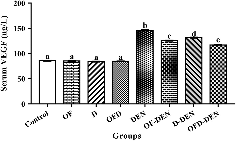
Fathy et al. 2017). Our study showed a significant increase (p < 0.05) in AFP and VEGF in the DEN-treated group. VEGF is known to play an essential role in tumor angiogenesis by inducing new vessel formation and promoting tumor invasion and metastasis (Xiang et al. 2011). In agreement with our results, it has long been recognized that the exposure of rats to certain carcinogens like DEN causes an increase in the circulating AFP levels that is associated with tumors (Song et al. 2013; Shahat et al. 2015).
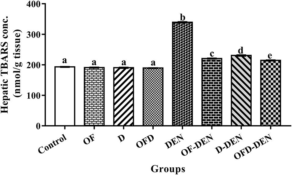
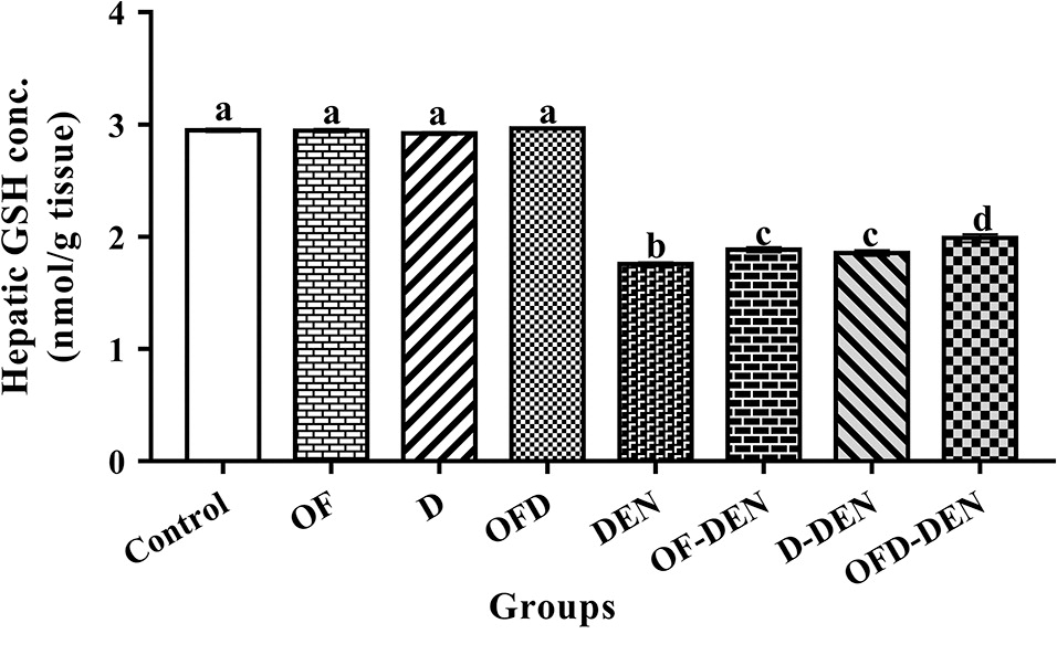
In our study, increased content of the lipid peroxidation products (i.e., TBARS) during DEN treatment provoked the generation of free radicals, which was confirmed by reduced oxidative stress markers of the antioxidant GSH and NO concentrations (Fathy et al. 2017). The significant decrease (p < 0.05) of NO in the liver tissue of the DEN-treated groups as compared with the control group might be the result of its interaction with superoxide to form peroxynitrite, which can react with cellular lipids, proteins, and DNA, and accelerates cell toxicity (Zahran et al. 2006; Bashandy et al. 2014). DEN induces oxidative stress possibly by the generation of reactive oxygen species (ROS), which are capable of initiating peroxidative damage to the cell. Lipid peroxidation (i.e., TBARS) is a useful marker of oxidative stress because it is correlated with increased production of ROS (Vasquez-Garzon et al. 2009). The increase of TBARS in the DEN-treated groups in our study reflects the enhancement of lipid peroxidation (Lykkesfeldt 2007; Fathy et al. 2017). In addition, this increase in tissue TBARS is considered an important marker of toxicity (Al-Saedi et al. 2015).
The lipid profile parameters showed a significant increase (p < 0.05) in TG, TC, LDL-C, and risk ratios in contrast to a significant decrease (p < 0.05) of HDL-C in the DEN-treated group. In line with the current data, free radicals impair liver functions and can be a major cause of hormonal imbalance. This imbalance induces hyperlipidemia through its multiple effects on lipid metabolism, including increased synthesis of cholesterol, TG, and LDL-C (Applebaum-Bowden et al. 1989). This indicates that DEN-induced oxidative stress might alter the hepatic lipid metabolism (Kim et al. 2006; Fathy et al. 2017); therefore, it is suggested that oxidative stress might be an important determinant of altered lipid metabolism from DEN treatment. Our results are also in accordance with those of Verna et al. (1996) and Tsukamoto et al. (2000) who reported an increase in lipids in the plasma of rats after DEN treatment, and attributed the hypercholesterolemia conditions to the stimulation of cholesterol synthesis in the liver after DEN treatment and its release from tissues due to destruction of cell membranes, and the mobilization of fats from the adipose tissues into the bloodstream in addition to mitochondrial dysfunction. Moreover, Bok et al. (1999) attributed hypercholesterolemia to increased activation of 3-Hydroxyl-3-methyl glutaryl coenzyme A (HMG-CoA) reductase enzyme, the key regulatory enzyme in the reduction of the overall process of cholesterol synthesis.
In accordance with Fathy et al. (2017), DEN treatment provoked oxidative stress that led to hematological disorders. Oxidative stress is evidenced by the significant decrease (p < 0.05) in the investigated hematological values (RBC count, Hb concentration, Hct percentage, platelet count, WBC count, and lymphocyte percentage) in contrast to a significant increase (p < 0.05) in neutrophil and monocyte percentage. Elsadek et al. (2017) reported an insignificant decrease in the hematological values in DEN-treated albino rats as compared with control rats. The longer period of our study might be the cause of the damage in the hematopoietic system or the cause of the increased permeability of cell membranes, which in turn caused osmotic swelling leading to erythrocyte hemolysis.
For decades, plants have been recognized to contain many natural antioxidants and the dietary intake of such antioxidants has played an important role in protection against free radicals and other diseases like cardiovascular diseases and cancer (Bao and Fenwick 2004). Our results showed that treatment of normal rats with OF and (or) D fruit extracts for 23 consecutive weeks did not produce any significant biochemical or hematological alterations as compared with the corresponding values in the control rats. Therefore, these natural antioxidant products are quite safe when administered to rats (Bashandy et al. 2014; Sheikh et al. 2014; Bashandy et al. 2016).
The present study showed that the two week pre-treatment of DEN-treated groups with OF and (or) D for 23 consecutive weeks significantly (p < 0.05) improved TBARS, GSH, and NO in the liver tissue as well as serum AFP, VEGF, lipid profile parameters, lipid risk ratios, and hematological values, in comparison with the corresponding values in the DEN-treated group. In addition, the OFD–DEN-treated group showed a greater improvement than the OF–DEN or D–DEN-treated groups revealing a synergistic effect. The effect of OFD may be attributed to antioxidant properties. This effect was evidenced by the ability of OFD to improve TBARS, GSH, and NO levels in the liver tissue homogenate as well as serum AFP and VEGF. In addition, the protective action of OF against lipid peroxidation, GSH, and NO concentration as factors modifying membrane organization might be related to the ability to scavenge the oxidation-initiating agents that are produced during the oxidation of proteins and lipids. The antioxidant effect of OF is mainly due to phenolic compounds, which are able to donate a hydrogen atom to free radicals, thus stopping the propagation chain reaction during the lipid peroxidation process (Azab and Nada 2004).
In light of the current data, evidence is accumulating to demonstrate that extra virgin olive oil is remarkably rich in effective phenolic antioxidants (at least 30 phenolic compounds) that could provide protection by inhibiting oxidative damage (Bashandy et al. 2014; Rubio et al. 2014). According to Tuck and Hayball (2002), the phenolic compounds present in extra virgin olive oil are strong antioxidants and radical scavengers. Olive and extra virgin olive oils contain a considerable amount of oleuropein, hydroxytyrosol, and tyrosol, which all strongly inhibit ROS production. However, lipid peroxidation and its chain reaction in LDL-C were interrupted if the LDL-C lipids were protected from free radicals by antioxidants like in the present study. Animal studies have suggested a protective effect of olive oil phenolics on LDL-C oxidation, and that it decreases lipid risk ratios (Visioli et al. 2000; Fito et al. 2005). Czerwinska et al. (2012) reported that oleacein, one of the main phenolic compounds present in olive oil, was a stronger inhibitor of neutrophil’s oxidative burst, and thus contributes to the cardiovascular health benefits of olive oil in the Mediterranean diet.
The fig results in our study are in accordance with Solomon et al. (2010) who attributed the antioxidant effect of figs to a very effective antioxidant component called cyanidin-3-rhamnoglucoside, which inhibits lipid peroxidation and reduces oxidative stress. The fig results are also in line with Lee et al. (2012) and Fathy (2014) who found that the administration of fig extract to irradiated rats led to improvements in the lipid profile, lipid risk ratios, and hematological parameters.
The date palm fruit extract results in our study are in agreement with previous reports (Ahmed et al. 2015; Bashandy et al. 2016). In addition, our results are in line with Saafi et al. (2011) who demonstrated that the pre-treatment of rats with date palm fruit extract restored liver damage induced by dimethoate, as revealed by the inhibition of hepatic lipid peroxidation. These results suggest that date palm fruit extract might be acting as a potent antioxidant. Orabi and Shawky (2014) concluded that date palm fruit possesses different pharmacological activities and has hemopoietic activity, as mercuric-chloride-treated rats supplied with date palm fruit extract showed a significant improvement in hematological parameters. Panahi and Asadi (2009) reported that the extract of date fruit was useful in controlling blood cholesterol levels and protecting against oxidative injury through the inhibition of LDL oxidation.
In conclusion, our results revealed that treatment with OF and (or) D fruit extracts resulted in improved biochemical and hematological parameters. In addition, the combined OFD treatments showed greater improvement revealing a synergistic effect producing a broad spectrum of antioxidative activities that create an effective defense system against free radical attack.

References

Ahmed AF, Al-Qahtani JH, Al-Yousef HM, Al-Said MS, Ashour AE, Al-Sohaibani M, et al. 2015. Proanthocyanidin-rich date seed extract protects against chemically induced hepatorenal toxicity. Journal of Medicinal Food, 18(3): 280–289.
Allain CC, Poon LS, Chan CS, Richmond W, and Fu PC. 1974. Enzymatic determination of total serum cholesterol. Clinical Chemistry, 20(4): 470–475.
Al-Qarawi AA, Abdel-Rahman H, Ali BH, Mousa HM, and El-Mougy SA. 2005. The ameliorative effect of dates (Phoenix dactylifera L.) on ethanol-induced gastric ulcer in rats. Journal of Ethnopharmacology, 98(3): 313–317.
Al-Saedi HF, Al-Zubaidy AA, Khattab YI, and Sahib HB. 2015. Effect of pentoxifylline against doxorubicin-induced nephrotoxicity in rabbits. International Journal of Pharmaceutical Sciences Review and Research, 30(1): 195–199 [online]: Available from globalresearchonline.net/journalcontents/v30-1/36.pdf.
Applebaum-Bowden D, McLean P, Steinmetz A, Fontana D, Matthys C, Warnick GR, et al. 1989. Lipoprotein, apolipoprotein and lipolytic enzyme changes following estrogen administration in post menopausal women. Journal of Lipid Research, 30: 1895–1906 [online]: Available from jlr.org/content/30/12/1895.abstract.
Azab K, and Nada A. 2004. Restitution of energy metabolism in irradiated rats considering curcumin antioxidant capacity and metal biotransformation. Arab Journal of Nuclear Sciences and Applications, 37(2): 169–176 [online]: Available from inis.iaea.org/search/searchsinglerecord.aspx?recordsFor=SingleRecord&RN=35089751.
Bao Y, and Fenwick GR. 2004. Future perspectives in phytochemical and health research. In Phytochemicals in health and disease. Edited by Y Bao, and RG Fenwick, eds. CRC Press, Boca Raton, Florida, USA. pp. 331–342 [online]: Available from taylorfrancis.com/books/e/9780203021408/chapters/10.1201%2F9780203021408-24.
Bashandy MA, Abd El- Rasheid HG, Hasan HF, and Fathy AH. 2014. Protective and therapeutic effects of olive oil and Ficus carica as natural antioxidants on some biochemical parameters in liver of γ-irradiated male albino rats. Al Azhar Bulletin of Science, 25(1): 1–16.
Bashandy MA, Abd-el-aal A, Ibrahim DF, and El-sharkawy MA. 2016. Protective effects of date palm extract as natural antioxidants on hepatotoxicity induced by Cerastes cerastes venom in albino rats. International Journal of Advanced Research, 4: 647–665 [online]: Available from journalijar.com/uploads/413_IJAR-9155.pdf.
Beutler E, Duron O, and Kelly BM. 1963. Improved method for the determination of blood glutathione. The Journal of Laboratory and Clinical Medicine, 61(5): 882–888.
Bok SH, Lee SH, Park YB, Bae KH, Son KH, Jeong TS, et al. 1999. Plasma and hepatic cholesterol and hepatic activities of 3-hydroxy-3-methyl-glutaryl-CoA reductase and acyl CoA: cholesterol transferase are lower in rats fed citrus peel extract or a mixture of citrus bioflavonoids. Journal of Nutrition, 129(6): 1182–1185.
Burstein M, Scholnick HR, and Morfin R. 1970. Rapid method for the isolation of lipoprotein from human serum by precipitation with polyanions. Journal of Lipid Research, 11: 583–595.
Czerwinska M, Kiss AK, and Naruszewicz M. 2012. A comparison of antioxidant activities of oleuropein and its dialdehydic derivative from olive oil, oleacein. Food Chemistry, 131(3): 940–947.
Elsadek B, Mansour A, Saleem T, Warnecke A, and Kratz F. 2017. The antitumor activity of a lactosaminated albumin conjugate of doxorubicin in a chemically induced hepatocellular carcinoma rat model compared to sorafenib. Digestive and Liver Disease, 49(2): 213–222.
Fathy AH. 2014. Influence of extracted Ficus and olive oil on some physiological and biochemical parameters in gamma irradiated albino rats. In Zoology. Al-Azhar University, Nasr city, Cairo, Egypt. 285 p.
Fathy AH, Bashandy MA, Bashandy SA, Mansour AM, and Elsadek B. 2017. Sequential analysis and staging of a diethylnitrosamine-induced hepatocellular carcinoma in male Wistar albino rat model. Canadian Journal of Physiology and Pharmacology, 95(12): 1462–1472.
Fito M, Cladellas M, de la Torre R, Marti J, Alcantara M, Pujadas-Bastardes M, et al. 2005. Antioxidant effect of virgin olive oil in patients with stable coronary heart disease: a randomized, crossover, controlled, clinical trial. Atherosclerosis, 181(1): 149–158.
Fossati P, and Prencipe L. 1982. Serum triglycerides determined colorimetrically with an enzyme that produces hydrogen-peroxide. Clinical Chemistry, 28(10): 2077–2080.
Gilani AH, Mehmood MH, Janbaz KH, Khan AU, and Saeed SA. 2008. Ethnopharmacological studies on antispasmodic and antiplatelet activities of Ficus carica. Journal of Ethnopharmacology, 119(1): 1–5.
IARC. 1987. Overall evaluations of carcinogenicity. IARC Monographs on the Evaluation of Carcinogenic Risk of Chemicals to Humans. International Agency for Research on Cancer, Lyon, France [online]: Available from monographs.iarc.fr/ENG/Monographs/suppl7/Suppl7.pdf.
Institute of Laboratory Animal Resources. 1996. Guide for the care and use of laboratory animals, eighth edition. Committee for the update of the guide and use of laboratory animals. National Research Council of the National Academies, National Academy Press, Washington, DC, USA [online]: Available from nap.edu/catalog/12910/guide-for-the-care-and-use-of-laboratory-animals-eighth.
Kim BJ, Hood BL, Aragon RA, Hardwick JP, Conrads TP, Veenstra TD, et al. 2006. Increased oxidation and degradation of cytosolic proteins in alcohol-exposed mouse liver and hepatoma cells. Proteomics, 6(4): 1250–1260.
Lee HY, Kim JH, Jeung HW, Lee CU, Kim DS, Li B, et al. 2012. Effects of Ficus carica paste on loperamide-induced constipation in rats. Food and Chemical Toxicology, 50(3–4): 895–902.
Lykkesfeldt J. 2007. Malondialdehyde as biomarker of oxidative damage to lipids caused by smoking. Clinica Chimica Acta, 380(1–2): 50–58.
Montgomery HA, and Dymock JF. 1961. The determination of nitrite in water. Analyst, 86: 414–416.
Orabi SH, and Shawky SM. 2014. Effect of date palm (Phoenix dactylifera) seeds extracts on hematological, biochemical parameters and some fertility indices in male rats. International Journal of Sciences: Basic and Applied Research (IJSBAR), 17(1): 137–147 [online]: Available from gssrr.org/index.php?journal=JournalOfBasicAndApplied&page=article&op=view&path%5B%5D=2446.
Panahi A, and Asadi M. 2009. Cholesterol lowering and protective effects of date fruit extract: an in vivo study. Toxicology Letters, 189: S235–S235.
Reagan-Shaw S, Nihal M, and Ahmad N. 2008. Dose translation from animal to human studies revisited. FASEB Journal, 22(3): 659–661.
Rubio L, Macia A, Castell-Auvi A, Pinent M, Blay MT, Ardevol A, et al. 2014. Effect of the co-occurring olive oil and thyme extracts on the phenolic bioaccessibility and bioavailability assessed by in vitro digestion and cell models. Food Chemistry, 149: 277–284.
Saafi EB, Louedi M, Elfeki A, Zakhama A, Najjar MF, Hammami M, et al. 2011. Protective effect of date palm fruit extract (Phoenix dactylifera L.) on dimethoate induced-oxidative stress in rat liver. Experimental and Toxicologic Pathology, 63(5): 433–441.
Shaarawy SM, Tohamy AA, and Elgendy SM. 2009. Protective effects of garlic and silymarin on NDEA-induced rats hepato-toxicity. International Journal of Biological Sciences, 5: 549–557.
Shahat AA, Alsaid MS, Kotob SE, and Ahmed HH. 2015. Significance of Rumex vesicarius as anticancer remedy against hepatocellular carcinoma: a proposal-based on experimental animal studies. Asian Pacific Journal of Cancer Prevention, 16(10): 4303–4310.
Sheikh BY, Elsaed WM, Samman AH, Sheikh BY, and Ladin AM. 2014. Ajwa dates as a protective agent against liver toxicity in rat. European Scientific Journal, 3: 358–368 [online]: Available from eujournal.org/index.php/esj/article/view/2957/2913.
Solomon A, Golubowicz S, Yablowicz Z, Grossman S, Bergman M, Gottlieb HE, et al. 2006. Antioxidant activities and anthocyanin content of fresh fruits of common fig (Ficus carica L.). Journal of Agricultural and Food Chemistry, 54(20): 7717–7723.
Solomon A, Golubowicz S, Yablowicz Z, Bergman M, Grossman S, Altman A, et al. 2010. Protection of fibroblasts (NIH-3T3) against oxidative damage by cyanidin-3-rhamnoglucoside isolated from fig fruits (Ficus carica L.). Journal of Agricultural and Food Chemistry, 58(11): 6660–6665.
Song Y, Jin SJ, Cui LH, Ji XJ, and Yang FG. 2013. Immunomodulatory effect of Stichopus japonicus acid mucopolysaccharide on experimental hepatocellular carcinoma in rats. Molecules, 18(6): 7179–7193.
Tricker AR, and Preussmann R. 1991. Carcinogenic N-nitrosamines in the diet: occurrence, formation, mechanisms and carcinogenic potential. Mutation Research, 259(3–4): 277–289.
Tsukamoto T, Inada K, Fukami H, Yamamoto M, Tanaka H, Kusakabe, et al. 2000. Mouse strain susceptibility to diethylnitrosamine induced hepatocarcinogenesis is cell autonomous whereas sex-susceptibility is due to the microenvironment: analysis with C3H↔BALB/c sexually chimeric mice. Japanese Journal of Cancer Research, 91: 665–673.
Tuck KL, and Hayball PJ. 2002. Major phenolic compounds in olive oil: metabolism and health effects. The Journal of Nutritional Biochemistry, 13(11): 636–644.
Vasquez-Garzon VR, Arellanes-Robledo J, and Garcia-Roman R. 2009. Inhibition of reactive oxygen species and pre-neoplastic le-sions by quercetin through an antioxidant defense mechanism. Free Radical Research, 43: 128–137.
Vayalil PK. 2002. Antioxidant and antimutagenic properties of aqueous extract of date fruit (Phoenix dactylifera L. Arecaceae). Journal of Agricultural and Food Chemistry, 50(3): 610–617.
Verna L, Whysner J, and Williams G. 1996. N-nitrosodiethylamine mechanistic data and risk assessment: bioactivation, DNA-adduct formation, mutagenicity, and tumor initiation. Pharmacology & Therapeutics, 71: 57–81.
Vinson JA, Zubik L, Bose P, Samman N, and Proch J. 2005. Dried fruits: excellent in vitro and in vivo antioxidants. Journal of the American College of Nutrition, 24(1): 44–50.
Visioli F, Caruso D, Galli C, Viappiani S, Galli G, and Sala A. 2000. Olive oils rich in natural catecholic phenols decrease isoprostane excretion in humans. Biochemical and Biophysical Research Communications, 278(3): 797–799.
Wieland H, and Seidel D. 1982. Improved assessment of plasma lipoprotein patterns. IV. Simple preparation of a lyophilized control serum containing intact human plasma lipoproteins. Clinical Chemistry, 28(6): 1335–1337.
Xiang Z, Zeng Z, and Fan J. 2011. Gene expression profiling of fixed tissues identified hypoxia-inducible factor-1α, VEGF, and matrix metalloproteinase-2 as biomarkers of lymph node metastasis in hepatocellular carcinoma. Clinical Cancer Research, 17: 5463–5472.
Yoshioka T, Kawada K, Shimada T, and Mori M. 1979. Lipid peroxidation in maternal and cord blood and protective mechanism against activated-oxygen toxicity in the blood. American Journal of Obstetrics and Gynecology, 135(3): 372–376.
Zahran AM, Azab KS, and Abbady MI. 2006. Modulatory role of allopurinol on xanthine oxidoreductase system and antioxidant status in irradiated rats. Egyptian Journal of Radiation Sciences and Applications, 19(2): 371–388 [online]: Available from inis.iaea.org/search/search.aspx?orig_q=RN:37071546.

Information & Authors

Information

Published In

cover image FACETS
FACETS
Volume 3Number 1October 2018
Pages: 584 - 597
Editor: Peter Zahradka

History

Received: 19 June 2017
Accepted: 14 March 2018
Version of record online: 28 May 2018

Data Availability Statement

All relevant data are within the paper.

Key Words

  1. blood count
  2. date palm fruit extract
  3. diethylnitrosamine
  4. fig fruit extract
  5. lipid profile
  6. olive oil

Sections

Subjects

Authors

Affiliations

Abdallah H. Fathy [email protected]
Department of Zoology, Faculty of Science, Al-Azhar University, Cairo 11651, Egypt
Experiments and Advanced Pharmaceutical Research Unit, Faculty of Pharmacy, Ain Shams University, Cairo 11566, Egypt
Mohamed A. Bashandy
Department of Zoology, Faculty of Science, Al-Azhar University, Cairo 11651, Egypt
Samir A. Bashandy
Department of Pharmacology, National Research Center, Cairo 12622, Egypt
Ahmed M. Mansour
Department of Pharmacology and Toxicology, Faculty of Pharmacy, Al-Azhar University, Cairo 11651, Egypt

Author Contributions

AHF, MAB, and AMM conceived and designed the study.
AHF, SAB, and AMM performed the experiments/collected the data.
AHF, MAB, and SAB analyzed and interpreted the data.
AHF and AMM contributed resources.
All drafted or revised the manuscript.

Competing Interests

The authors have declared that no competing interests exist.

Metrics & Citations

Metrics

Other Metrics

Citations

Cite As

Export Citations

If you have the appropriate software installed, you can download article citation data to the citation manager of your choice. Simply select your manager software from the list below and click Download.

Cited by

1. Extracts of Jordanian Date Palm Fruit (Phoenix Dactylifera L.) Inhibit Human Mammary Adenocarcinoma (MCF-7) Cells In Vitro by Inducing Cell Viability
2. Synergistic effects of Ficus Carica extract and extra virgin olive oil against oxidative injury, cytokine liberation, and inflammation mediated by 5-Fluorouracil in cardiac and renal tissues of male albino rats
3. Hepatotoxicity and renal toxicity induced by gamma-radiation and the modulatory protective effect of Ficus carica in male albino rats
4. The beneficial effect of natural antioxidants from olive oil with fig and date palm fruit extracts on biochemical and hematological parameters in rats treated with doxorubicin and γ-radiation

View Options

View options

PDF

View PDF

Get Access

Media

Media

Other

Tables

Share Options

Share

Share the article link

Share on social media